Picture this: you capture a microscope image of a whole mouse brain and want to share the results with colleagues. The problem is, it’s 10 gigabytes—far too large to share via email or a free image sharing tool—not to mention download.
Sharing images is a common challenge in the microscopy world, and it’s one of the many obstacles our OlyVIA software can solve.
Today, we’ll explore how OlyVIA software works and show you different ways to leverage this free tool to simplify your image sharing process.
What is OlyVIA software?
Simply put, OlyVIA is our free image viewer software that can read data captured by our SLIDEVIEW VS200 research slide scanner, as well as our FV3000 series confocal microscopes. With our optional Net Image Server (NIS) solution, customers can securely and quickly store, review, and share confocal images and virtual slides on the web database.
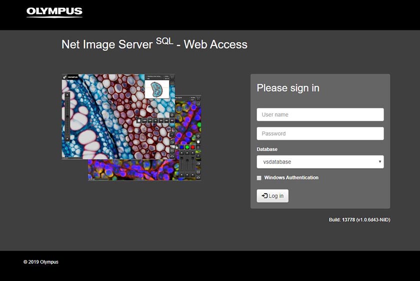
OlyVIA web log-on page
Here are three OlyVIA features that our customers like the most.
1. Image sharing
OlyVIA is an invaluable digital assistant to help keep everyone on the same page—and that’s why customers love using this software for image sharing in educational training and scientific collaboration.
Consider this example: in the classroom, a professor can add a link in the curriculum to an area of interest on a slide image. Students can then click the link, log on, and view this image at any time to complete their homework.
In addition, educators can present the digital slides on a large monitor, so all students can simultaneously look at an image. By doing so, educators can build up student interest in using the microscope and facilitate group discussion.
In the workplace, OlyVIA software is often used to train and collaborate with personnel.
2. Macro-to-micro viewing
A second reason customers like using OlyVIA software is the ability to go from the big picture to fine details in seconds.
A good analogy is that OlyVIA software works similarly to your favorite navigation app on your smartphone. Just like you can see an entire town and then zoom down into street view, OlyVIA enables you to view a mouse brain and then zoom closer to look at the individual neurons. You can see how this works in the image below.
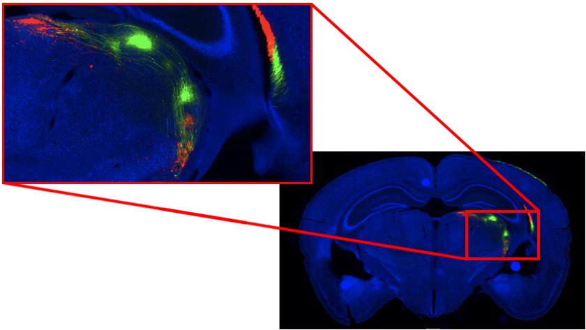
By quickly going between tissue and cellular levels in a single image, you can perform efficient quantitative data analyses.
3. Intuitive user interface
Another reason that customers love OlyVIA software is the easy-to-use and powerful user interface (UI). Here are four notable features:
- Slide annotation: Write notes directly on important areas of a digital slide.
- Touch screen: Using a touch screen capable device, you can easily navigate OlyVIA. Rotate, zoom in and out, or move up and down using your fingers. Alternatively, you can use a computer mouse to perform the same tasks.
- Search bar: Quickly find and access folders, annotations, and images in the search bar.
- Channels: Visualize biomarkers by selecting different fluorescence channels. This is a handy tool for many clinical research applications, like immunotherapy.
Now that you know a few favorite features, let’s explore two of OlyVIA’s lesser known but powerful functions.
1. Side-by-side digital slide comparisons.
One lesser known feature of OlyVIA software is the ability to perform side-by-side image comparisons.
It works like this: select two or more images, zero in on the same location, and hit the Compare button. This simultaneous review feature is especially helpful for biomarker validation and analyzing serial section tissue morphology.
2. Importing documents.
You may not realize that OlyVIA isn’t limited to just digital images. With the new SLIDEVIEW VS200 research slide scanner, you can now import and view documents, such as your lab notes, along with your images.
Now you can access all your notes and images in one place to improve your research workflow.
How to set up OlyVIA
Ready to try out these features for yourself? You can access OlyVIA in two convenient ways:
- OlyVIA via web: To use OlyVIA on the web, you need our optional Net Image Server (NIS) solution and ISS web server. You can create a web interface to make images accessible via an Internet browser that supports HTML5, such as Google Chrome and Mozilla Firefox.
- OlyVIA via app: Download the free OlyVIA Desktop app for Windows PCs by visiting our software downloads page and selecting Research Slide Scanner.