The human eye is qualitative—not quantitative. Don’t believe us? Then take a look at the two squares below. Which one is larger?

Most people would choose the one on right. Yet, the two squares are the same size. This is a simple example of an optical illusion, and it teaches us how the human eye is not good at measuring something quantitatively.
Despite this, many cell culture processes today still rely on visual observation. This post will explore the challenges that visual observation introduces into the cell culture process and share the importance of moving to a quantitative approach.
To begin, let’s walk through the standard cell culture process.
The Standard Cell Culture Process
In a standard cell culture workflow, researchers start seeding a specific number of cells based on the information from cell counting. Next, they culture cells on a dish or flask and put them into an incubator to make the cells grow. Every two or three days, they take out the cell culture vessel from the incubator to visually check the cell condition under a microscope.
Most people check cell morphology and confluency since they are important factors of cell health:
- Cell morphology tells you if cells are generally healthy.
- Confluency is a key factor that can determine whether you do a passage. If cells are overconfluent, cells can become quiescent or die. Historically, researchers check cell confluency using visual observation under a microscope.
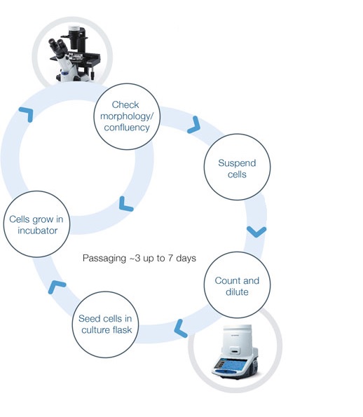
Figure 1. Standard workflow for the cell culture process.
Now remember what we saw in the optical illusion example. We know that human eyes are not as quantitative as we may think. In many cases, 70% confluency under visual observation is not actually 70%. It might be 80% or maybe 60%.
Also, cell confluency will be estimated differently by each person who performs a cell culture check. Since human eye-based estimation is unstable, it can affect the quality of your cell culture and lead to inconsistent results.
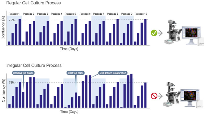
Figure 2. (Top) optimal and (bottom) irregular cell culture process.
The Standard Workflow after Cell Culture
After the cell culture process, you might move on to microscopy imaging to explore their biological behavior, to cell-based assays to assess compounds, or to cell production on a larger scale (Figure 3). But without quantitative cell culture, you can run into issues.
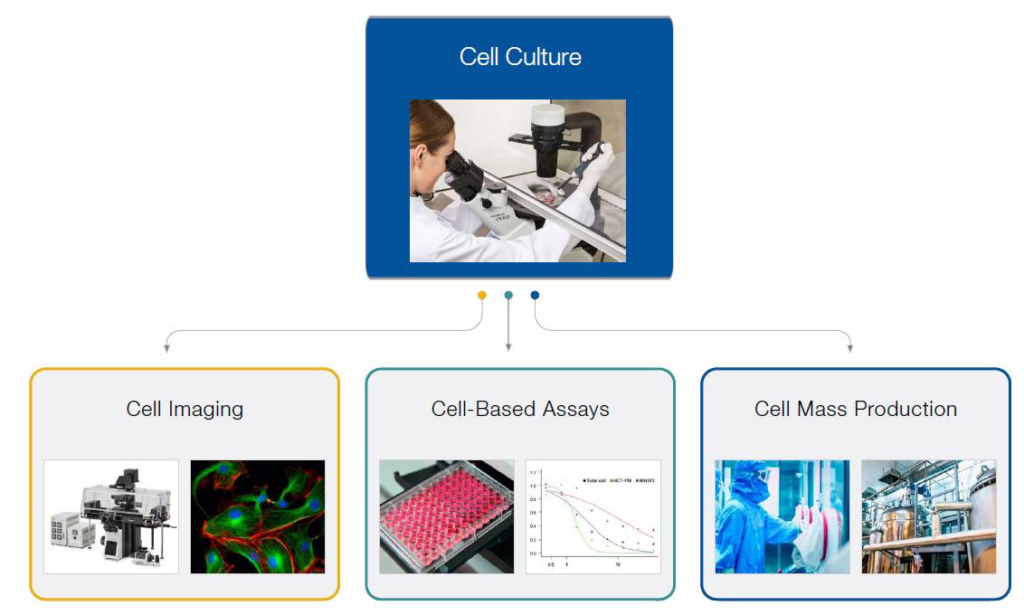
Figure 3. Typical workflow after cell culture.
Consider this example: cell-based assays evaluate drug efficacy using cultured cells. Yet, if the quality of the cell culture is inconsistent, the result of the assay will be unreliable because the cells could react differently to a drug candidate.
The importance of quantitative cell culture is also clearly mentioned in the Assay Guidance Manual published by the National Center for Advancing Translational Sciences (NCATS).
The manual states, "Standardizing primary cell culture conditions is essential for robust assay performance. For experiments where cells are used from a new source (patient or animal) for each experiment, responsiveness will vary, and separate normalizations will be required for each experiment."
Another section reads, "Particular attention to cell culture conditions is critical for the success of the assay. Cells should be confluent, but not overgrown, for the assay to work properly."
As you can see, the quality of your cell culture affects your process after cell culture. Just consider the lost time and added expenses that can occur if you notice your results are unreliable after the experiment.
It’s clear that taking a quantitative approach to cell culture is critical for improving your process—but how do you actually get more consistent results? It all comes down to using the right tools.
Helpful Tools for Quantitative Cell Culture
While you can’t rely on the human eye for quantitative cell culture results, advanced image analysis technologies are making up for it.
With breakthrough image analysis technology using artificial intelligence, it has become much easier to analyze label-free images, such as this phase contrast image of cells shown below (Figure 4).
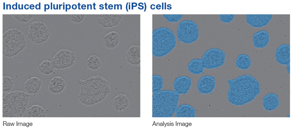
Figure 4. Example of a machine-learning-based cell analysis.
Combining a microscopy image with AI-driven analysis technology makes it possible to estimate cell confluency using machine learning. Since machine learning can use stable and standardized parameters, it offers a more robust and quantitative method than traditional visual evaluation with a microscope.
This powerful technology is employed in our CM20 incubation monitor and confluency checker software, providing you with reproducible and quantitative data for successful experiments.
Related Content
Video: CM20 Incubation Monitoring System
Efficiently Analyze the Cell Growth Characteristics of an Autologous Chondrocyte Culture
Quantifying Confluency of iPS Cell Colonies after Derivation