Not Available in Your Country
Sorry, this page is not
available in your country.
Vue d’ensemble
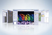
 | Fonctionnement intuitif. Processus très simple.La plateforme CellSens d’Olympus vous permet de contrôler entièrement l’affichage et l’emplacement des icônes, des barres d’outils et des commandes. Vous pouvez donc faire évoluer et adapter le logiciel en fonction de vos besoins de recherche en constante évolution. |
|---|
cellSens Packages |
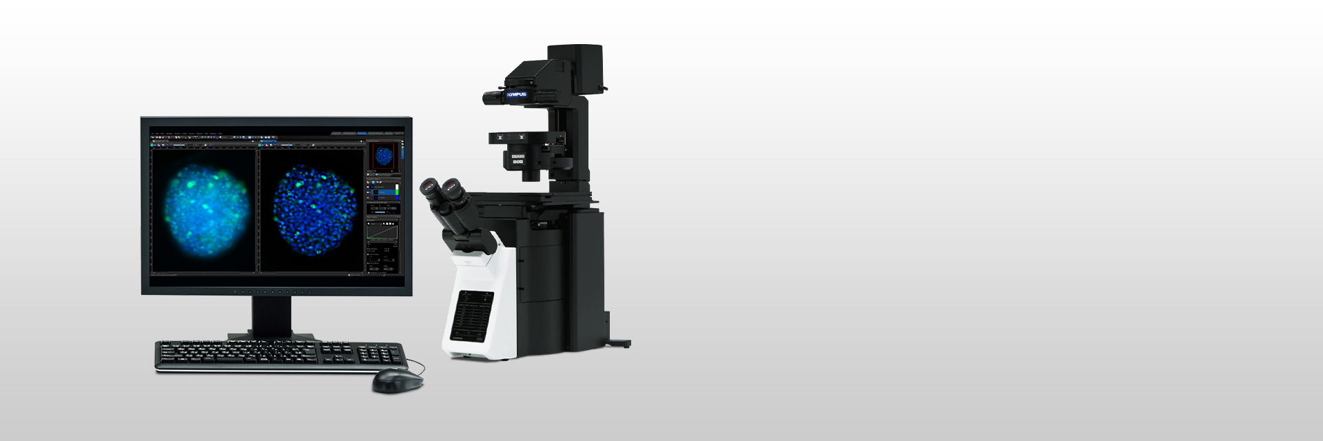
cellSens EntrycellSens Entry est la pierre angulaire idéale pour les chercheurs qui souhaitent passer à l’acquisition et à la documentation d’images numériques leur offrant tous les outils nécessaires pour acquérir des images de façon simple. |
*cellSens Entry n’est pas offert dans certaines régions. |
cellSens StandardLe progiciel cellSens Standard d’Olympus est fondé sur le progiciel cellSensEntry et permet l’acquisition d’images multiples au moyen de processus de saisie d’image avancés (p. ex. capture à intervalles) et du contrôle des composants de microscopes motorisés et codés. |
|
cellSens DimensioncellSens Dimension est le progiciel le plus polyvalent de la gamme cellSens d’Olympus puisqu’il est doté d’une fonction d’acquisition d’images entièrement automatisée, de puissants outils d’analyse et plus encore. |
|
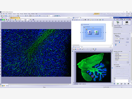 | Acquisition d’images en 5DAcquérez des images en cinq dimensions à l’aide de divers outils, comme le Graphic Experiment Manager (GEM) et le Well Navigator qui vous aident à visualiser de façon conviviale votre acquisition de données. |
|---|
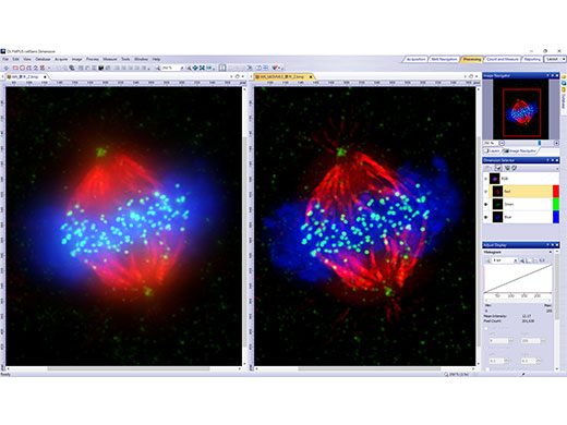
Traitement et partage d’imagesVoyez les données réelles dans vos images grâce à la déconvolution TruSight et à d’autres techniques de traitement d’images. Partagez facilement vos résultats au moyen du mode de conférence, ou glissez-déposez des données dans les rapports préconfigurés. |
|
|---|
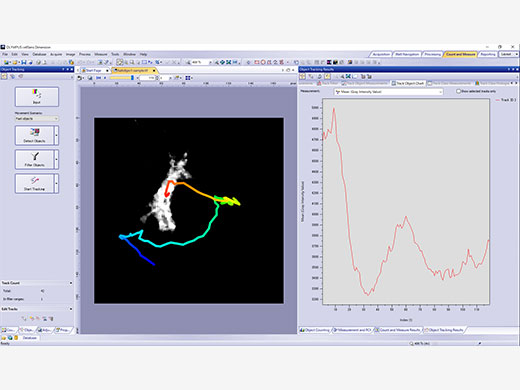
| Outils d’analyse puissantsTravaillez de façon dynamique avec vos images pour en extraire le plus de données possibles et obtenir des résultats expérimentaux fiables. La technologie d’apprentissage profond du logiciel (TruAI) offre une analyse par segmentation améliorée. Servez-vous du gestionnaire de macros « Macro Manager » pour automatiser des flux de travail complets de l’analyse jusqu’à la sauvegarde des images. |
|---|
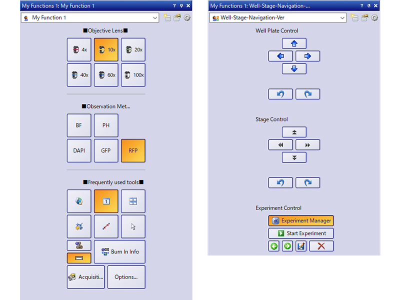
Interface utilisateur personnalisableChoisissez une disposition recommandée pour l’acquisition d’images et l’analyse, ou créez votre propre disposition en utilisant l’ensemble d’outils My Functions. |
|
|---|
Besoin d’assistance? |
Le logiciel cellSens n'est pas destiné à un usage diagnostique. |
Caractéristiques
cellSens Functions and Optional Solutions |
| Dimension | Standard | Entry | |||
|---|---|---|---|---|---|
| Layout | User experience customization | ✓ | ✓ | ✓ | |
| View | Overlay multiple images | ✓ | ✓ | - | |
| Document groups for side-by-side image comparison | ✓ | ✓ | ✓ | ||
| Movie playback | ✓ | ✓ | ✓ | ||
| Tile view (multiple images in a single data set shown side-by-side) | ✓ | ✓ | ✓ | ||
| Slice view for orthogonal plane viewing of 3D or time-lapse data sets | ✓ | - | - | ||
| Voxel viewer for isosurface and volumetric rendering of 3D and 4D data sets | ✓ | - | - | ||
| Image Acquisition | Snap/movie acquisition | ✓ | ✓ | ✓ | |
| Time-lapse at specified interval | ✓ | ✓ | - | ||
| Automated multiwavelength | ✓ | ✓ | - | ||
| Z-stack | ✓ | - | - | ||
| Multidimensional (XYZT and wavelength) | ✓ | - | - | ||
| Graphical Experiment Manager | ✓ | - | - | ||
| Manual panoramic imaging (Instant MIA and Manual MIA) | ✓ | Manual Process | Manual Process | ||
| Multiposition visitation and stage navigator | Multiposition | - | - | ||
| Automated panoramic imaging (auto MIA, requires motorized stage) | Multiposition | - | - | ||
| Instantly create EFI image (manual or motorized Z) | ✓ | Manual Process | Manual Process | ||
| Simultaneous multicolor Imaging (requires two identical cameras** or image splitter) | ✓ | - | - | ||
| Live deblurring | ✓ | - | - | ||
| High dynamic range imaging (HDRI) | ✓ | - | - | ||
| Multiwell plate acquisition | Well plate navigator and Multiposition | - | - | ||
| Image Processing | Geometry/combine/filter processing | ✓ | ✓ | - | |
| Fluorescence unmixing | ✓ | - | - | ||
| Brightfield unmixing | Count & Measure | - | - | ||
| Deblurring (No/Nearest Neighbor, Wiener Filter) | ✓ | - | - | ||
| Kymograph | ✓ | - | - | ||
| 2D deconvolution | ✓ | - | - | ||
| 3D deconvolution (constrained iterative deconvolution with GPU process) | CI Deconvolution | - | - | ||
| Image Analysis | Phase analysis | ✓ | - | - | |
| Object measurements and classification | Count & Measure | Count & Measure | - | ||
| Interactive 2D measurements | ✓ | ✓ | ✓* | ||
| Intensity plot over time/z | ✓ | - | - | ||
| Colocalization | ✓ | - | - | ||
| Object counting (manual) | ✓ | ✓ | - | ||
| Object tracking | Tracking and Count & Measure | - | - | ||
| Online ratio and kinetics | Ratio/FRET | - | - | ||
| Ratio analysis (offline) | ✓ | - | - | ||
| FRET analysis | Ratio/FRET or Life Science Analysis | - | - | ||
| FRAP analysis | Photo Manipulation or Life Science Analysis | - | - | ||
| Cell count and confluency measurements | ✓ | Confluency Checker | - | ||
| Deep Learning | Training of Neural Networks | Deep Learning | Deep Learning | - | |
| Inference using trained Neural Networks (offline/online) | Deep Learning or Count & Measure | Deep Learning or Count & Measure | - | ||
| Documentation and Collaboration | Automatically compose MS Word reports | ✓ | - | - | |
| Database image and data management solution for microscopy | Database Core | Database Core | - | ||
| Open database and load records/documents from database | Database Client | Database Client | Database Client | ||
| Remoting | Remote live image viewing | NetCam | NetCam | - | |
|
* Three points angle, four points angle, arbitrary line, closed polygon, polyline and perpendicular line only. Interactive 2D measurements option is needed to add other measurement tools and make exporting Excel spreadsheets possible.
** Supported cameras: iXon Ultra 897, Zyla 5.5 (USB 3.0), Zyla 4.2 (USB 3.0/CamLink), Neo, iXon Ultra 888, ImagEM X2, ORCA-Flash 4.0 (V2/V3), Prime 95B, Prime BSI, Prime BSI Express, Sona4.2B-11, ORCA Fusion, ORCS-Fusion BT, ORCA-QUEST |
| Dimension | Standard | |||
|---|---|---|---|---|
| Layout | User experience customization | ✓ | ✓ | |
| Microscope Control | Microscope Control | ✓ | ✓ | |
| View | Slice view for orthogonal plane viewing of 3D or time-lapse data sets | ✓ | - | |
| Voxel viewer for isosurface and volumetric rendering of 3D and 4D data sets | ✓ | - | ||
| Image Acquisition | Automated multiwavelength | ✓ | ✓ | |
| Z-stack | ✓ | - | ||
| Multidimensional (XYZT and wavelength) | ✓ | - | ||
| Instantly create EFI image (manual or motorized Z) | ✓ | Manual Process | ||
| Automated panoramic imaging (auto MIA, requires motorized stage) | Multiposition | - | ||
| Manual panoramic imaging (Instant MIA and Manual MIA) | ✓ | Manual Process | ||
| Simultaneous multicolor imaging (requires two identical cameras or image splitter)*1 | ✓ | - | ||
| Live deblurring | ✓ | - | ||
| High dynamic range imaging (HDR) | ✓ | - | ||
| Multiwell plate acquisition | Well Plate Navigator and Multiposition | - | ||
| Image Processing | MIA | ✓ | ✓ | |
| Geometry/combine/filter processing | ✓ | ✓ | ||
| Morphological filter | Count & Measure | Count & Measure | ||
| Fluorescence unmixing | ✓ | - | ||
| Brightfield unmixing | Count & Measure | - | ||
| Kymograph | ✓ | - | ||
| 2D deconvolution | ✓ | - | ||
| 3D deconvolution (constrained iterative deconvolution) | CI Deconvolution | - | ||
| Image Analysis | Interactive 2D measurements | ✓ | ✓ | |
| Object counting (manual) | ✓ | ✓ | ||
| Colocalization | ✓ | - | ||
| Object measurements and classification | Count & Measure | Count & Measure | ||
| Object tracking | Tracking and Count & Measure | - | ||
| Online ratio and kinetics | Ratio/FRET | - | ||
| Ratio analys (offline) | ✓ | - | ||
| FRET analysis | Ratio/FRET or Life Science Analysis | - | ||
| FRAP analysis | Life Science Analysis | - | ||
| Cell count and confluency measurements | ✓ | Confluency Checker | ||
| Deep Learning | Training of Neural Networks | Deep Learning | Deep Learning | |
| Inference using trained Neural Networks (offline/online) | Deep Learning or Count & Measure | Deep Learning or Count & Measure | ||
| Report | Report function (Microsoft Word is needed) | ✓ | - | |
| Documentation and Collaboration | Database image and data management solution for microscopy | Database Core | Database Core | |
| Open database and load records/documents from database | Database Client | Database Client | ||
* Supported cameras: iXon3 897, Zyla 5.5 (USB 3.0), Zyla 4.2 (USB 3.0/CamLink), Neo, iXon Ultra 888, ImagEM X2, ORCA-Flash 4.0 (V2/V3), Prime 95B, Prime BSI, Prime BSI Express, Sona4.2B-11, ORCA-Fusion, ORCA-Fusion BT, ORCA-QUEST |
Products with Confirmed Functionality |
| Dimension | Standard | Entry | |||
|---|---|---|---|---|---|
| Olympus | Camera | DP22, DP23, DP23M, DP27, DP28, DP73, DP74, DP75, DP80, XM10, XC10, XC30, XC50, UC30, UC50, UC90, LC20, LC30, LC35, SC50, SC100, SC180 | ✓ | ✓ | ✓ |
| Micoscope | BX43, BX53, BX63, BX61, BX61WI, IX83, IX73, IX81, SZX16A | ✓ | ✓ | - | |
| IX81-ZDC, IX81-ZDC2 | ✓ | - | - | ||
| Peripherals | BX-DSU, IX3-DSU, IX3-ZDC, IX3-ZDC2, IX2-DSU, IX2-ZDC, IX2ZDC2, U-CBF, cellTIRF (multiline, single line), MT20, USB-ODB converter, Real Time Controller (U-RTCE), U-FCB | ✓ | - | - | |
| Light Source | U-LGPS | ✓ | ✓ | - | |
| Hamamatsu | Camera | ImagEMX2, ORCA-Flash 4.0 V2, ORCA-Flash 4.0 V3, ORCA-Flash 4.0 LT, ORCA-Flash 4.0 LT PLUS, ORCA-Flash 4.0 LT3, ORCA-Fusion, ORCA-Fusion BT, ORCA-QUEST | ✓ | - | - |
| ORCA-spark | ✓ | ✓ | - | ||
| Image Splitter | W-View Gemini | ✓ | - | - | |
| Q-Imaging | Camera | Retiga 6000 | ✓ | - | - |
| Photometrics | Camera | Prime (PCI-Express), Prime 95B, Prime BSI, Prime BSI Express, Moment | ✓ | - | - |
| Image Splitter | Dual View DV2 / QuadView QV2 | ✓ | - | - | |
| Andor | Camera | iXon X3 897, iXon Ultra 897, iXon Ultra 888, iXon Life 888, iXon Life 897, Sona4.2B-11,Zyla4.2/Zyla4.2 PLUS (Camera-link,USB3.0), Zyla5.5 (Camera-link 10tap,USB3.0), ZL41 Cell 4.2 (Camera-link,USB3.0), Neo5.5 | ✓ | - | - |
| Vincent Associates | Shutter | Uniblitz shutter (VCM-D1, VMM-D1, VMM-D3) | ✓ | ✓ | - |
| CoolLED | Light Source | pE-1, pE-2, pE800, pE-4000 | ✓ | - | - |
| pE-300white, pE-300ultra, pE-340fura | ✓ | ✓ | - | ||
| Excelitas | Light Source | X-Cite120LED, X-Cite XYLIS, X-Cite TURBO | ✓ | - | - |
| Lumencor | Light Source | SOLA SEII, SEII 365, Spectra X | ✓ | - | - |
| Sutter | Shutter, FW | Lambda 10-3/10-B | ✓ | - | - |
| Prior | Motorized XY Stage | ProScan III, Optiscan III | Multiposition | - | - |
| Shutter, FW, Z-drive | ProScan (I, II, III) , Optiscan III | ✓ | - | - | |
| Piezo Z (Control via Real Time Controller) | NanoScanZ NZ100 | ✓ | - | - | |
| Ludl | Motorized XY Stage | Mac 6000 | Multiposition | - | - |
| Shutter, FW, Z-drive | Mac 6000 | ✓ | - | - | |
| Märzhäuser | Motorized XY Stage | Tango, Pilot Stage | Multiposition | - | - |
Z-drive Controller | Tango | ✓ | - | - | |
| Physik Instrumente | Piezo Z (Control via Real Time Controller) | PIFOC P-721 | ✓ | - | - |
| Applied Scientific Instrumetation | Motorized XY Stage | MS-2000 | Multiposition | - | - |
| Z-drive Controller | MS-2000 | ✓ | - | - | |
| National Instruments | Digital TTL Device | NI USB-6501, NI USB-6343 | ✓ | - | - |
| Yokogawa | CSU | CSU-X1, CSU-W1 | ✓ | - | - |
| Regarding the detailed Windows OS compatibility, please contact an Evident sales representative. |
| Dimension | Standard | |||
|---|---|---|---|---|
| Olympus | Camera | DP22, DP23, DP23M, DP27, DP28, DP73, DP74, DP75, DP80 | ✓ | ✓ |
| Micoscope | BX43, BX53, BX63, BX61, BX61WI, IX83, IX73, IX81, SZX16A | ✓ | ✓ | |
| IX81-ZDC, IX81-ZDC2 | ✓ | - | ||
| Peripherals | BX-DSU, IX3-DSU, IX3-ZDC, IX3-ZDC2, IX2-DSU, IX2-ZDC, IX2ZDC2, U-CBF, Real Time Controller (U-RTCE) | ✓ | - | |
| Light Source | U-LGPS | ✓ | ✓ | |
| Hamamatsu | Camera | ImagEMX2, ORCA-Flash 4.0 V2, ORCA-Flash 4.0 V3, ORCA-Flash 4.0 LT PLUS, ORCA-Fuision, ORCA-Fusion BT, ORCA-QUEST | ✓ | - |
| ORCA-spark | ✓ | ✓ | ||
| Image Splitter | W-View Gemini | ✓ | - | |
| Q-Imaging | Camera | Retiga 6000 | ✓ | - |
| Photometrics | Camera | Prime, Prime 95B, Prime BSI, Prime BSI Express, Moment | ✓ | - |
| Image Splitter | Dual View DV2 /QuadView QV2 | ✓ | - | |
| Andor | Camera | iXon X3 897, iXon Ultra 897, iXon Ultra 888, iXon Life 888, iXon Life 897, Sona4.2B-11, Zyla4.2/Zyla4.2 PLUS (Camera-link,USB3.0), Zyla5.5 (Camera-link 10tap,USB3.0), ZL41 Cell 4.2 (Camera-link,USB3.0), Neo5.5 | ✓ | - |
| Vincent Associates | Shutter | Uniblitz shutter (VCM-D1, VMM-D1, VMM-D3) | ✓ | ✓ |
| Ludl | Motorized XY Stage | Mac 6000 | Multiposition | - |
| Shutter, FW, Z-drive | Mac 6000 | ✓ | - | |
| Prior | Motorized XY Stage | ProScan III, Optiscan III | Multiposition | - |
| CoolLED | Light Source | pE-1, pE-2, pE800, pE-4000 | ✓ | - |
| pE-300white, pE-300ultra, pE-340fura | ✓ | ✓ | ||
| Excelitas | Light Source | X-Cite120LED, X-Cite XYLIS, X-Cite TURBO | ✓ | - |
| Lumencor | Light Source | SOLA SEII, SEII 365, Spectra X | ✓ | - |
| Sutter | Shutter, FW | Lambda 10-3/10-B | ✓ | - |
| National Instruments | Digital TTL Device | NI USB-6501, NI USB-6343 | ✓ | - |
| Yokogawa | CSU | CSU-W1 | ✓ | - |
| Regarding the detailed Windows OS compatibility, please contact an Evident sales representative. |
Compatible image formats |
| Read and write | JPEG, JPEG2000, TIFF, BMP, AVI, PNG, VSI, PSD(Adobe Photoshop), Big TIFF, OIR | ||||
|---|---|---|---|---|---|
| Read only | GIF, OIF/OIB(FLUOVIEW format), Cell, STK (MetaMorph), MRC (Medical Research Council) | ||||
System requirements |
| OS | Microsoft Windows 10 Pro (64-bit(21H2 build 19044.1466), Microsoft Windows 11 Pro(64-bit)(22H2) | ||||
|---|---|---|---|---|---|
| OS Language | English, Simplified Chinese, Japanese, German and Italian (Entry and Standard) | ||||
| CPU | Intel Core i5, Intel Core i7, Intel Xeon Recommended for high-speed image acquisition: QuadCore | ||||
| RAM |
8GB for general applications, 16GB or more is recommended for high-speed image acquisition, 32GB or more is recommended for Deep learning
(For DP23/DP28/DP23M, dual memory is recommended for high frame rate imaging) | ||||
| HDD |
5 GB for installation
Recommended for high speed image acquisition: Solid State Drive (SSD) | ||||
Software version update
Version update is available for the next version following the version written on license card. (Exclude updating sub-minor versions) An update that spans 2 or more major or minor versions is required an update license.
|
Ressources
Notes d'application
Blogue
Documentation
Documents infographiques
Manuel
Vidéos
Webinaire
Téléchargements
Installers and Version Checker |
cellSens V4.2.1 64bit InstallercellSens V4.2 64bit Installer | cellSens V4.1.1 64bit Installer | cellSens V3.2 64bit Installer |
cellSens V2.3 32bit InstallercellSens V2.3 64bit Installer | cellSens V1.18 32bit InstallercellSens V1.18 64bit Installer | cellSens V1.16 32bit InstallercellSens V1.16 64bit Installer |
VERSION 1.7 OR LATER | Upgrading to a Windows 10 PC | |
Notes de mise à jour
Version 4.2.1New hardware support
New function/improvement
Version 4.2New hardware support
New function/improvement
Version 4.1.1New hardware support
Minor bug improvements
Version 4.1New hardware support
New function/improvement
Version 3.2New hardware support
New function/improvement
Version 2.3New hardware support
New function/improvement
Version 2.2New hardware support
New function/improvement
Version 2.1New hardware support
Product portfolio changes
New function/improvement
|


![cellSens [ver.4.2.1] User Manual](https://static5.olympus-lifescience.com/modules/imageresizer/64e/8c7/4132b5b5e5/112x84p74x50.jpg)
![cellSens [ver.4.2.1] Installation Manual](https://static2.olympus-lifescience.com/modules/imageresizer/99b/1ba/44507be555/112x84p63x50.jpg)
![cellSens [ver.4.2.1] Hardware Manual](https://static3.olympus-lifescience.com/modules/imageresizer/241/054/8e949a7a55/112x84p69x50.jpg)
![cellSens [ver.4.2.1] Database Manual](https://static5.olympus-lifescience.com/modules/imageresizer/254/161/ab9aae4bef/112x84p67x50.jpg)
![cellSens [ver.4.2] Database Manual](https://static4.olympus-lifescience.com/modules/imageresizer/051/014/b73c664ebb/112x84p63x50.jpg)
![cellSens [ver.4.2] Installation Manual](https://static5.olympus-lifescience.com/modules/imageresizer/1e2/f4e/59fde3cca6/112x84p69x50.jpg)
![cellSens [ver.4.2] Hardware Manual](https://static3.olympus-lifescience.com/modules/imageresizer/ed0/00b/da9613f82e/112x84p67x50.jpg)
![cellSens [ver.4.2] User Manual](https://static3.olympus-lifescience.com/modules/imageresizer/31c/7b4/c7022777f0/112x84p71x50.jpg)
![cellSens [ver.4.1] Installation Manual](https://static4.olympus-lifescience.com/modules/imageresizer/d67/038/32f7f54871/112x84p61x50.jpg)
![cellSens [ver.4.1] Hardware Manual](https://static3.olympus-lifescience.com/modules/imageresizer/626/e28/7713ca10bb/112x84p68x50.jpg)

![cellSens [ver.4.1] Database Manual](https://static2.olympus-lifescience.com/modules/imageresizer/a4d/346/5e7903657a/112x84p63x50.jpg)
![cellSens [ver.4.1] User Manual](https://static5.olympus-lifescience.com/modules/imageresizer/15a/70a/01d83866c2/112x84p61x50.jpg)


