
Ask the Experts
Ask the Expertsは、ライフサイエンス研究や顕微鏡の活用法に関する質問にお答えするポータルページです。当社及び協力機関・企業のエキスパートによるライブウェビナーに参加して最新トピックを学んだり、質問や課題をリアルタイムで問い合わせたりすることができます。見逃し配信にも対応しているので、配信後は「オンデマンドウェビナー」から繰り返しご視聴いただけます。もし興味のあるトピックが見つからない場合は、お問い合わせフォームからご要望いただくか、当社スタッフに直接ご連絡ください。
ライブウェビナー | オンデマンドウェビナー | エキスパートのご紹介
Now You Have the Power to See More
Experts

The Olympus VS200 digital slide scanner has been very well received since its release in March 2020. With a reliable, flexible, and customizable design, the system has been adopted by various industries including research, geology, and many others. View this session to find out more and see examples of samples scanned using this popular addition to the Olympus product range.
The Use of Multiplexing in Microscopy for Better Understanding the Skin Immune System in the Context of the Tissue
Experts

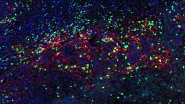
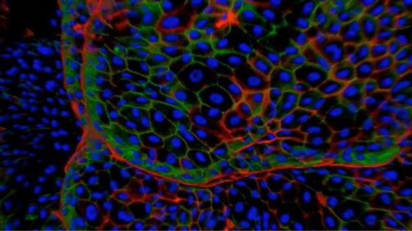
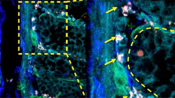
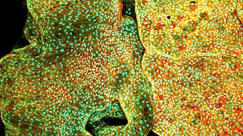
The skin is the first line of defense and the immune system’s biggest barrier for combating pathogens. Being able to accurately characterize and identify immune cell subtypes, tissues structures, and cell distribution in the skin under steady-state conditions provides a powerful tool for understanding the first immunological strategies and biological processes that occur in the presence of pathogens. In this webinar we will review technical aspects involved in the experimental process and explore how complementary imaging technologies might assist us to better understand the immune system.
The presentation is divided into three parts. First, an introduction of the Hugh Green Cytometry Centre will be presented and an overview of the histology and bioimaging technological platforms available. Second, the multiplexing methodology will be discussed, where several topics need to be considered for the design and development of a successful polychromatic panel for microscopy. Finally, preliminary results from a research project will be presented that constitutes part of a diploma program from The Royal Microscopical Society. The project focuses on the identification of immune cell types in the whole mount skin in relation to tissues structures (e.g., blood vessels and lymphatic network). It also centers on the immune cells’ distribution in the tissue as a first barrier of defense against pathogens.
Hyperspectral and Brightfield Imaging Combined with Deep Learning Uncover Hidden Regularities of Colors and Patterns in Cells and Tissues
Experts

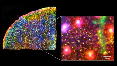
The Australian Research Council Centre of Excellence for Nanoscale Biophotonics draws on key advances of the 21st century, nanoscience, and photonics to help understand life at the molecular level. In this presentation, next-generation technologies developed in our Centre for probing, imaging, and interacting with the living systems will be discussed. These address the key challenges of ultrasensitive detection of key analytes in real complex environments and molecular complexity, and they support both novel therapies and diagnostics.
Create a Smarter Cell Culture Workflow
Experts

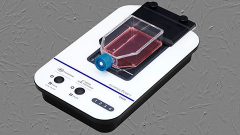
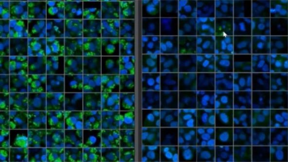
In this webinar, expert Joanna Hawryluk explores how the OLYMPUS Provi™ CM20 incubation monitoring system can help improve the health and stability of cell cultures through machine learning. With the aid of AI, the CM20 monitor automatically measures cell conditions using constant analysis parameters to provide quantitative data—all while your cultures remain safely in the incubator.
Culture and Quantitative 3D Imaging of Organoids: Challenges and Solutions
Experts

Dr. Anne Beghin
Assistant Professor, Research Mechanobiology Institute
National University of Singapore
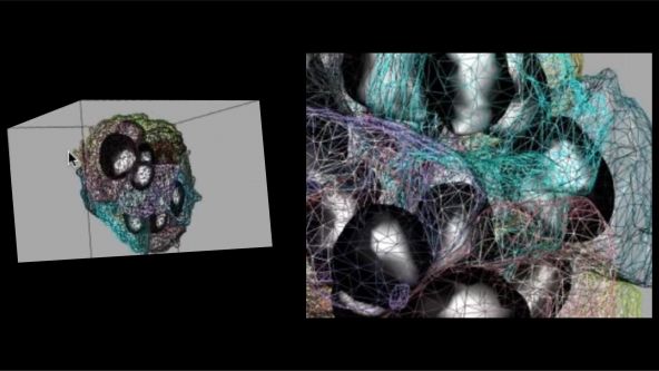
Turning organoids into impactful translational models includes being able to culture them and assess those that develop robustly with physiologically relevant architecture. However, quantitative comparisons and statistical analysis at high content, which are mandatory to describe the complexity of such multicellular 3D objects are not possible owing to the lack of high-throughput 3D imaging methods. We have thus engineered a versatile High Content Screening (HCS) device to streamline all the steps of organoid culture to exploit its potential in morphogenesis understanding. Our approach comprises a new generation of versatile scaffolding cell culture multiwell chips with embedded optical components (= lighting JeWells) that enables rapid 3D imaging.
Converting from 2D to 3D: Bio-Techne Solutions for Your 3D Culture
Experts

Organoid and three-dimensional (3D) cell culture are emerging as pivotal systems for understanding human organ development, modeling disease, screening for drug efficacy or toxicity, and investigating personalized medicine. Usually they are derived from primary tissue, embryonic stem cells (ESCs), or induced pluripotent stem cells (iPSCs), which are capable of self-renewal and differentiation.
Utilizing Tumoroids to Explore Anti-Tumor Immunity in Rectal Cancer
Experts

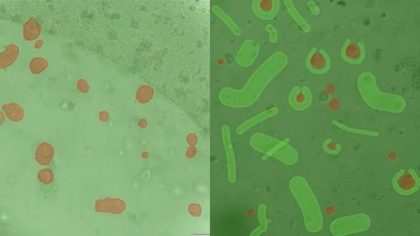
Globally colorectal cancer is a significant public health burden. It is the third most commonly diagnosed cancer and fourth leading cause of cancer related deaths in the world. A subset of patients diagnosed with rectal cancer require neoadjuvant chemoradiotherapy (NACRT) prior to surgery. However, there is a spectrum of response to this therapy with only 10-20% of patients achieving a complete pathological response. In addition, 20-40% of patients will demonstrate no response to this treatment. There is currently no method that predicts how a patient will respond to NACRT accurately. In order to investigate the mechanisms underpinning how patients respond to therapy, patient derived tumouroids have been utilised. These personalised in vitro three-dimensional tumour models recapitulate the in vivo tumour of origin genotypically. The Ramsay laboratory (Peter MacCallum, Melbourne) has successfully co-cultured patient-matched rectal cancer tumouroids with tumour infiltrating lymphocytes (TILS) in a novel in vitro assay, preliminary data generated suggests this assay has the ability to predict the response of a patient to treatment with NACRT prior to instigation of neo-adjuvant therapy. This assay provides a pre-clinical platform that encapsulates the hosts immune response toward their tumour. However, manual analysis of the data generated from this assay is time consuming and limits the clinical utility of this platform. Machine-based learning to develop artificial neural networks capable of analysing data produced from the killing assay has been developed to automate analysis. Automated analysis utilising artificial neural networks is a feasible approach to expedite the processing of data generated from the cytotoxic killing assays and will improve the clinical utility of this platform to direct personalised patient therapy.
Tissue Optical Clearing Imaging: From In Vitro to In Vivo
Experts

Dr. Dan Zhu
Professor
Wuhan National Laboratory for Optoelectronics, Huazhong University of Science and Technology, Wuhan, China
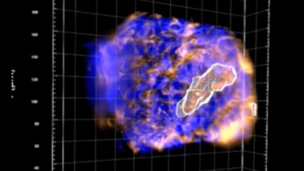
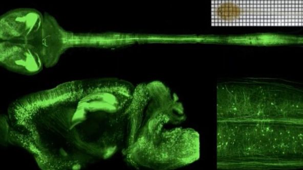
Biomedical optical Imaging, as a powerful tool has been applied for observing biomedical tissue structural and functional information with high resolution and contrast unattainable by any other method. However, the high scattering of turbid biological tissues limits the penetration of light, leading to strongly decreased imaging resolution and contrast as light propagates deeper into the tissue. Fortunately, novel tissue optical clearing technique provide a way for reducing the scattering of tissue and improving the optical imaging quality. This presentation will introduce our progress from in vitro and in vivo of tissue optical clearing imaging, including developing in vitro optical clearing methods, such as FDISCO and MACS. Meanwhile, we will also demonstrate in vivo skull/skin optical clearing window for imaging structural and functional of cutaneous / cortical vascular and cells, also manipulating cortical vasculature.
3D Microscopy: Understanding the Give and Take on Instrument Performance to Enable Informed Decisions
Experts

Biologists have a significant toolbox at their disposal when it comes to microscopically imaging 3D samples, such as organoids. From widefield microscopy to confocal, superresolution, multiphoton and lightsheet, each have their own set of pros and cons that must be carefully considered before making an informed choice on the most suitable to address your biological question. Often a correlative approach is required, applying several techniques to address the question from different perspectives. It is also crucial to consider the method of sample preparation and optimise each of the potential steps which can include fixation, permeabilisation, labelling and mounting. Further, the images generated by all techniques can be enhanced with post-processing techniques, such as deconvolution, which can enable or help to improve subsequent image analysis and interpretation.
Advances in 3D Optical Imaging Technologies: An Overview
Experts

With rapid development in fluorescent proteins, synthetic fluorochromes, and digital imaging, advanced 3D imaging technologies are now available to investigators to provide critical insights into the fundamental nature of cellular and tissue functions. 3D and 4D imaging systems have become very common tools among biologists. However, there are several technical challenges and limitations in performing successful 3D and 4D imaging. Olympus has developed a wide range of 3D imaging microscopes to overcome these challenges and to satisfy the requirements of researchers across different disciplines.
Investigating Spheroid Architecture Using the FV3000 Confocal Microscope
Experts

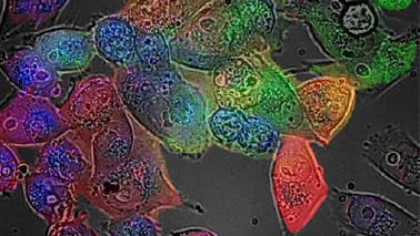
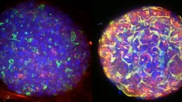
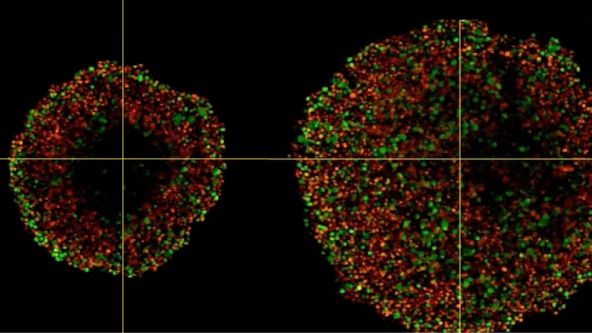
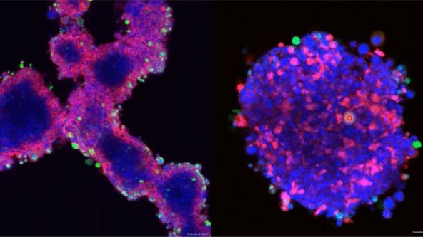
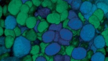
Phenotypic tumour heterogeneity arising due to differentially cycling cell populations has been implicated in increased therapy resistance. This phenomenon cannot be assessed in adherent cell culture, where microenvironmental conditions are homogeneous. Thus, we utilise melanoma spheroids to model the 3D tumour microenvironment including the extracellular matrix (ECM) and study spheroid structure, necrotic region, individual cell arrangement within and gene expression patterns. We achieve this by exploiting the fluorescence ubiquitination cell cycle indicator (FUCCI) system to monitor cell cycle stages as a surrogate marker for phenotypic tumour heterogeneity, tissue clearing and confocal microscopy using FV3000.
Prostate Cell Lineage Hierarchy and Plasticity
Experts

Dr. Dong Gao
Principal Investigator
Shanghai Institute of Biochemistry and Cell Biology, Chinese Academy of Sciences
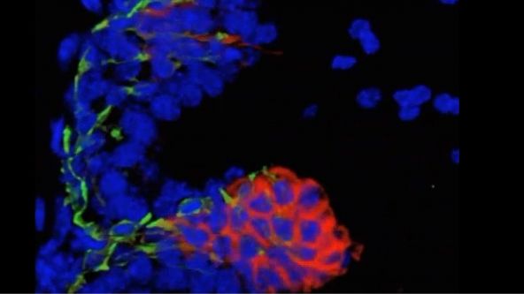
Prostate cancer is one of the most common cancers worldwide and also the second leading cause of cancer-related death in males in Western countries. Although the majority of human primary prostate cancers have a luminal phenotype, both basal cells and luminal cells can serve as cellular origins of prostate cancer in model systems. However, the stem cell-like plasticity of defined prostate epithelial cells and the cellular origin of prostate cancer under physiological conditions have not been identified. Recently, prostate basal and luminal cell populations were both shown to be self-sustaining, and both cell types could initiate prostate cancer. However, the oncogenic transformation of basal cells requires basal to luminal cell transition. In addition, luminal cells were shown to have greater tendency to be the cells of origin for prostate cancer in some contexts.
3D Segmentation for Fluorescence Images: From Qualitative to Quantitative
Experts

Dr. Yu Weimiao
Head of Computational & Molecular Pathology Lab (CMPL) Agency of Science
Technology and Research
Cells are 3D functional elements in biology science and they are actively moving to perform their functions. Collective cell migration is appreciated as an important model for the understanding of the mechanism governing the cell movement in Vivo and in Vitro. It is a highly kinetic process involved in immune response, wound healing, tissue development and cancer metastasis. Recent decades have seen the fast development of various optical imaging techniques with excellent spatial-temporal resolution, dimensionality and scale. The generation of novel probes have also allowed us to acquire the movies of migrating cells with specific proteins/molecules. However, we lack of advanced solution to analyse such high-content and highly dynamic images/videos.
An In Vitro System for Evaluating Anticancer Drugs Using Patient-Derived Tumor Organoids
Experts

Dr. Motoki Takagi
Professor
Medical-Industrial Translational Research Center, Fukushima Medical University
Patient-derived tumor organoids (PDOs) represent a promising preclinical cancer model that better replicates disease, compared with traditional cell culture models. We have established a novel series of patient-derived tumor organoids (PDOs) from various types of tumor tissues from the Fukushima Translational Research Project, which are designated as Fukushima (F)-PDOs. F-PDOs could be cultured for >6 months and formed cell clusters with similar morphologies to their source tumors. Comparative histological and comprehensive gene-expression analyses also demonstrated that the characteristics of PDOs were similar to those of their source tumors, even following long-term expansion in culture. In addition, suitable high-throughput assay systems were constructed for each F-PDO in 96- and 384-well plate formats.
Study the Function of Stromal Cells through Intestinal Organoid Co-Culture Technology
Experts

Dr. Ningbo Wu
Associate Professor
Shanghai Institute of Immunology, Shanghai Jiao Tong University School of Medicine
For a long period of time, intestinal mesenchymal stromal cells have been considered as a relatively simple and homogeneous group of cells. With the help of single cell transcriptomics studies, it has now been clear that these cells are quite complex and heterogeneous. However, the detailed cellular and molecular mechanisms that regulate the function of these cells remains poorly understood. Therefore, the ability to perturb and evaluate the function of these stromal cells is critical to the understanding of intestinal stem cell niche and the etiology of the inflammatory bowel diseases and colitis associated colorectal cancer.
NoviSight™ Demonstration: 3D Image Analysis and Statistical Software for Organoids and Spheroids
Experts

Three-dimensional cell culture models such as patient-derived organoids (PDO) and spheroids have increased in popularity because they can provide a 3D microenvironment that more closely reproduces in vivo conditions compared to 2D monolayer culture. Phenotypic and functional heterogeneity arise among cancer cells within the same tumor because of genetic change, environmental differences and reversible changes in cell properties. Therefore, evaluation of cell-specific responses is important for accurate prediction of drug efficacy and kinetics in vivo.
Olympus Organoid Conference 2021
During the Olympus Organoid Conference, cell biologists, microscopists, and image analysis experts shared their insights and answered questions on the latest developments in organoid technologies.
Depth Matters: Transforming Biology for More Realistic and Meaningful Pursuits
Experts

Dr. Gowri Balachander
Research Fellow, Translation Mechanobiology lab, National University of Singapore
Improvements to in vitro three-dimensional (3D) models are making them increasingly better at mimicking in vivo-like cellular behavior. Every tissue presents a distinct microenvironment with a unique blend of biochemical and biophysical components that dictate cellular behavior. Recreation of critical features of tissues that nurtures recapitulation of in vivo-like cellular behavior is the essence of an effective 3D model. In this webinar, through specific examples of 3D models for tissue development and cancer, we will revisit the fundamental principles of designing 3D models that can effectively recapitulate critical features of the tissues in vitro and applications of such models in mechanistic studies and drug testing. Our work also highlights the importance of 3D imaging systems, such as laser scanning confocal microscopes, which are necessary for such work.
患者由来のオルガノイドとスフェロイドの三次元高スループット画像解析
Experts
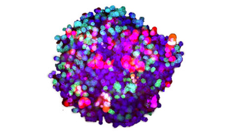
オルガノイドとスフェロイドがin vivo条件を忠実に再現できる一方で、イメージングに基づく解析では、高い空間分解能により細胞固有の反応をモニタリングできます。 オリンパスでは患者由来のがんのオルガノイドとスフェロイドを使用して、イメージングに基づく三次元解析および医薬品評価方法を開発しています。
.jpg?rev=1940)
デジタル画像処理パート2:先進の画像処理フィルター
Experts

このトピックについてお届けした最初のウェビナーでは、光学顕微鏡で取得した画像がサンプルを完全に表すものではないことを学びました。 誤差の原因は常に存在し、誤差を最小限に抑えることしかできないので、多くの場合は最終的な画像解析の前に、イメージング実験のローデータに何らかのデジタル画像処理を施す必要があります。
ICSI—Past, Present & Future
Experts

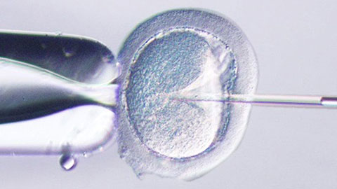
In this webinar, Dr. Pai will be sharing on what’s new in the management of male infertility with special focus on Intra Cytoplasmic Sperm Injection (ICSI). He will be describing the latest diagnostic modalities in male infertility especially sperm DNA fragmentation, genetic testing, cryotozoospermia and microsurgical TESA in cases of non-instructive azoospermia.
.jpg?rev=3E0D)
顕微鏡対物レンズ:魔法が起こる場所
Experts


このウェビナーでは、LaurenとKlausが顕微鏡などの複雑なシステムにおける優れた光学製品の重要性にクローズアップしていきます。 最終的な画質にとって重要なさまざまな要素について説明します。
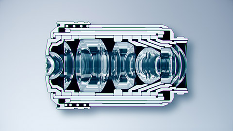
Microscope Objectives—Where the Magic Happens
Experts


In this webinar, Olympus microscope experts Ganesh and Wei Juan discuss the importance of good optics in a complex microscope system and what optical features are important for high final image quality.
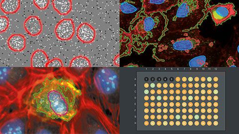
ハイコンテントスクリーニング:カスタム解析を簡単に
Experts


このウェビナーでは、ディープラーニングとハイコンテントスクリーニング分野の当社専門家であるManoelと尚平が、scanRシステムのアッセイビルダーを紹介します。 scanRシステムは、独自のサンプルナビゲーション方法と、フローサイトメトリーから発想を得た解析を特長とする、オリンパスのハイコンテントスクリーニング専用プラットフォームです。