Evident LogoOlympus Logo

Ask the Experts

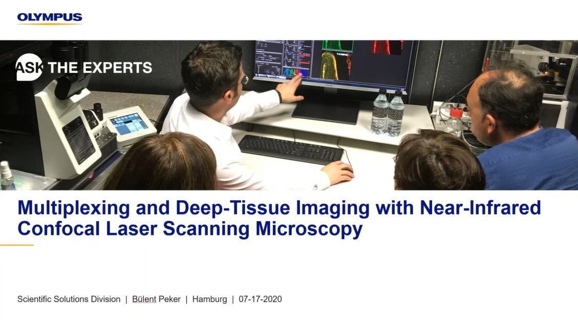
Multiplexing and Deep Tissue Imaging with Near-Infrared Confocal Laser Scanning Microscopy

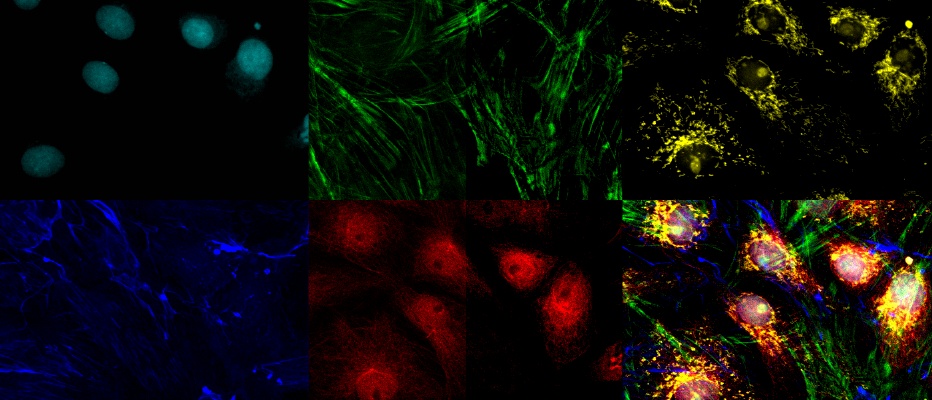
In this webinar, you’ll learn about fluorescence multiplexing and deep tissue imaging using near-infrared (NIR) laser light. NIR laser sources can help in visualizing biological structures more clearly and at higher resolution deep within a specimen. NIR excitation can also enable the use of more fluorescent dyes without spectral overlap.

Presenters:

Bülent Peker, Senior Product Marketing Manager
Rebecca Bonfig, Product Manager, Confocal Microscopy

FAQ

Webinar FAQs | Near-infrared (NIR) laser microscopy

Do you lose detection of lower wavelengths when using near infrared (NIR) imaging?

Due to the detectors’ design, you retain full availability to detect wavelengths starting at 400 nm. Lower wavelength emissions can still be collected at that range. We still have our standard and GaAsP photomultiplier tubes (PMTs) as well, so NIR is more like an add-on.

What are the benefits of using NIR over two-photon microscopy?

Two-photon microscopy is typically used for high-depth imaging in the red part of the spectrum. NIR offers an easy entry point into this application in a cost-effective way. With NIR, you can use confocal microscopes, so you don’t need to enter into a different class of microscopy system. As well as being much less expensive, NIR also offers greater multiplexing and spectral capabilities.

What is the VPH grating used for inside the microscope?

Volume-phase holographic (VPH) grating is part of our TruSpectral technology in standard and GaAsP detectors. It is used to generate the spectrum of light for spectral imaging. The advantage of VPH grating is that it is much more light efficient than using other reflection-type spectral gratings.

Do I have to purchase NIR lasers with the system or can I upgrade later?

One of the great features of this system is its modularity, so it can be upgraded at any time to add NIR lasers and detectors.

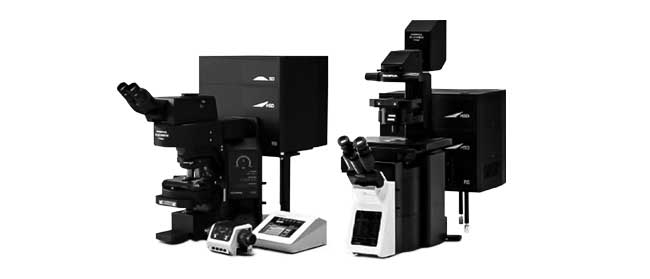
Is the six-detector mode available on both the IX and BX series?

Yes, the six-detector mode is available on both the IX series of inverted microscopes and the BX series of upright microscopes.

Does the FV3000 support NIR excitation wavelengths other than 730 and 785 nm?

The 730 nm and 785 nm laser lines are carefully selected as a result of cooperation with a specialized manufacturer that develops high-quality, stable lasers at these wavelengths.

However, there is a laser port available for the NIR range, so customization is possible. We encourage conversations to discuss the possibility of using different laser lines. The FV3000 microscope’s beam combiner makes it highly amenable to customization, and the system has optical coatings that support wavelengths out to 1600 nm, so users also have the ability to route their own NIR lasers into the system.

Can you image 730 and 785 nm on the same image set?

It is currently not possible to image both 730 nm and 785 nm, but we are always working to improve the system.

What are the differences between GaAsP detectors and competitors’ newest detectors?

We have not compared the systems side by side, but it’s important to remember that the detector is just one part of the whole system sensitivity. Make sure to look at the whole optical train and attached optics and how well they do in the NIR range. We know that there are several aspects during the light path that increase light efficiency in the NIR range, and it is also important that the optics are suited for the NIR range.

Can you use a white light laser with the FV3000 microscope?

White light lasers (WLLs) are not available on the FV3000 system. Olympus has a broad range of laser lines, which allow us to adapt the system for the needs of individual users and match the laser to their applications. There is also a cost-effectiveness factor and the advantage of the whole system not needing to be down if there is a problem with the WLL. Instead, you have individual laser lines that are affordable and easy to change.


関連製品

共焦点レーザー走査型顕微鏡

FV3000

  • ガルバノスキャナーのFV3000と、ガルバノ/レゾナントのハイブリッドキャナーのFV3000RS
  • 高感度・高精度のTruSpectral分光検出器
  • 新開発マルチチャンネルモードによる16チャンネルのアンミキシング
  • 様々なアプリケーションやサンプルの種類に応じて倒立と正立の組み合わせから選択可能

Multiplexing and Deep Tissue Imaging with Near-Infrared Confocal Laser Scanning Microscopy

In this webinar, you’ll learn about fluorescence multiplexing and deep tissue imaging using near-infrared (NIR) laser light. NIR laser sources can help in visualizing biological structures more clearly and at higher resolution deep within a specimen. NIR excitation can also enable the use of more fluorescent dyes without spectral overlap.

FAQ

Webinar FAQs | Near-infrared (NIR) laser microscopy

Do you lose detection of lower wavelengths when using near infrared (NIR) imaging?

Due to the detectors’ design, you retain full availability to detect wavelengths starting at 400 nm. Lower wavelength emissions can still be collected at that range. We still have our standard and GaAsP photomultiplier tubes (PMTs) as well, so NIR is more like an add-on.

What are the benefits of using NIR over two-photon microscopy?

Two-photon microscopy is typically used for high-depth imaging in the red part of the spectrum. NIR offers an easy entry point into this application in a cost-effective way. With NIR, you can use confocal microscopes, so you don’t need to enter into a different class of microscopy system. As well as being much less expensive, NIR also offers greater multiplexing and spectral capabilities.

What is the VPH grating used for inside the microscope?

Volume-phase holographic (VPH) grating is part of our TruSpectral technology in standard and GaAsP detectors. It is used to generate the spectrum of light for spectral imaging. The advantage of VPH grating is that it is much more light efficient than using other reflection-type spectral gratings.

Do I have to purchase NIR lasers with the system or can I upgrade later?

One of the great features of this system is its modularity, so it can be upgraded at any time to add NIR lasers and detectors.

Is the six-detector mode available on both the IX and BX series?

Yes, the six-detector mode is available on both the IX series of inverted microscopes and the BX series of upright microscopes.

Does the FV3000 support NIR excitation wavelengths other than 730 and 785 nm?

The 730 nm and 785 nm laser lines are carefully selected as a result of cooperation with a specialized manufacturer that develops high-quality, stable lasers at these wavelengths.

However, there is a laser port available for the NIR range, so customization is possible. We encourage conversations to discuss the possibility of using different laser lines. The FV3000 microscope’s beam combiner makes it highly amenable to customization, and the system has optical coatings that support wavelengths out to 1600 nm, so users also have the ability to route their own NIR lasers into the system.

Can you image 730 and 785 nm on the same image set?

It is currently not possible to image both 730 nm and 785 nm, but we are always working to improve the system.

What are the differences between GaAsP detectors and competitors’ newest detectors?

We have not compared the systems side by side, but it’s important to remember that the detector is just one part of the whole system sensitivity. Make sure to look at the whole optical train and attached optics and how well they do in the NIR range. We know that there are several aspects during the light path that increase light efficiency in the NIR range, and it is also important that the optics are suited for the NIR range.

Can you use a white light laser with the FV3000 microscope?

White light lasers (WLLs) are not available on the FV3000 system. Olympus has a broad range of laser lines, which allow us to adapt the system for the needs of individual users and match the laser to their applications. There is also a cost-effectiveness factor and the advantage of the whole system not needing to be down if there is a problem with the WLL. Instead, you have individual laser lines that are affordable and easy to change.


関連製品

共焦点レーザー走査型顕微鏡

FV3000

  • ガルバノスキャナーのFV3000と、ガルバノ/レゾナントのハイブリッドキャナーのFV3000RS
  • 高感度・高精度のTruSpectral分光検出器
  • 新開発マルチチャンネルモードによる16チャンネルのアンミキシング
  • 様々なアプリケーションやサンプルの種類に応じて倒立と正立の組み合わせから選択可能
Experts
Bülent Peker
Senior Product Marketing Manager
Evident

こんにちは。Bülent Pekerと申します。レーザー走査型検鏡法の専門家として任命されました。私が検鏡法と光工学に初めて興味を持ったのは、時間分解2光子検鏡法を扱っていた物理化学の博士課程の頃で、この情熱は今もずっと続いています。

オリンパスに13年以上勤務し、当社の最先端のレーザー走査型顕微鏡を市場に送り出すために、チームを手伝ってきました。特に、多光子システムのアプリケーションとレーザー走査システムのカスタム化の可能性に興味を駆り立てられています。 

Rebecca Bonfig
Product Manager, Confocal Microscopy

こんにちは。Rebecca Bonfigと申します。オリンパスで共焦点検鏡法のプロダクトマネージャーをしています。University of Louisvilleの生理学・生物物理学部で副甲状腺ホルモンによる腎リン酸輸送体Npt2aの転写後調節について研究し、大学院を修了しました。2015年からオリンパスに在籍し、米国、カナダ、中南米で、FLUOVIEW製品のサポートを担当しています。

Multiplexing and Deep Tissue Imaging with Near-Infrared Confocal Laser Scanning Microscopy2024年4月20日
このページはお住まいの地域ではご覧いただくことはできません。
Ask the Expert Sign-up

By clicking subscribe you are agreeing to our privacy policy which can be found here.

このページはお住まいの地域ではご覧いただくことはできません。

このページはお住まいの地域ではご覧いただくことはできません。