A cell contains anywhere from one to several thousand mitochondria, depending on the metabolic requirements of the organism. For example, a heart muscle cell requires thousands of mitochondria to feed its rigorous energy needs whereas an epithelial tissue cell may only need a fraction of that amount. These large organelles are found in almost all eukaryotes: plants, animals, protists, and even fungi, and were first discovered with a light microscope in the 1800s. Their grainy appearance under the microscope inspired the name mito-chondria, originating from the Greek words for "thread" and "granules" respectively. Our present understanding of mitochondrial behavior began when a method for isolating the organelles without damaging them was developed around the 1950s. In the digital video presented above, human osteosarcoma epithelial cells (U2Os line) are expressing an enhanced yellow fluorescent protein (EYFP) fusion to a mitochondrial targeting signal.
Video 1 - Run Time: 66 Seconds
Video 2 - Run Time: 57 Seconds
Video 3 - Run Time: 66 Seconds
Video 4 - Run Time: 67 Seconds
Video 5 - Run Time: 66 Seconds
Video 6 - Run Time: 62 Seconds
Video 7 - Run Time: 66 Seconds
Video 8 - Run Time: 66 Seconds
Video 9 - Run Time: 67 Seconds
Because it possesses its own circular DNA, and reproduces independently of the cell in which it is found, the mitochondrion is different from most other organelles; an apparent case of endosymbiosis. Scientists hypothesize that millions of years ago small, free-living prokaryotes were engulfed, but not consumed, by larger prokaryotes, perhaps because they were able to resist the digestive enzymes of the host organism. The two organisms developed a symbiotic relationship over time, the larger organism providing the smaller with ample nutrients and the smaller organism providing ATP molecules to the larger one. Eventually, according to this view, the larger organism developed into the eukaryotic cell and the smaller organism into the mitochondrion. In the digital video presented above, human osteosarcoma epithelial cells (U2Os line) are expressing an enhanced yellow fluorescent protein (EYFP) fusion to a mitochondrial targeting signal.
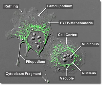
A mitochondrion’s elaborate structure is very important to the function of the organelle. Two specialized membranes encircle each mitochondrion, dividing the organelle into a narrow intermembrane space and a much larger internal matrix, each of which contains highly specialized proteins. The outer membrane of a mitochondrion contains many channels formed by the protein porin and acts like a sieve, filtering out molecules that are not needed. Similarly, the inner membrane, which is highly convoluted so that a large number of infoldings called cristae are formed, also allows only certain molecules to pass through it and is much more selective than the outer membrane. To make certain that only those materials essential to the matrix are allowed into it, the inner membrane utilizes a group of transport proteins that will only transport the correct molecules. Together, the various compartments of a mitochondrion are able to work in harmony to generate ATP in a complex multi-step process. In the digital video presented above, human osteosarcoma epithelial cells (U2Os line) are expressing an enhanced yellow fluorescent protein (EYFP) fusion to a mitochondrial targeting signal.


