전문가에게 문의하기
생명 과학 현미경에 대해 문의할 수 있는 원스톱 포털인 전문가에게 문의하기에 오신 것을 환영합니다. 라이브 웹 세미나 전문가들과 함께 최신 주제에 대해 토론하거나 언제든지 전문가에게 문의하여 질문과 과제를 해결하십시오. 웹 세미나를 놓치셨습니까? 문제 없이 다시 들으실 수 있습니다. 토론하고 싶으신 주제가 없습니까? 피드백 양식을 사용하거나 전문가에게 직접 문의하여 도움을 요청하십시오.
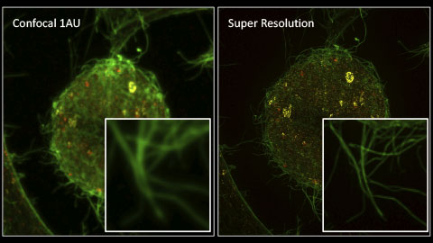
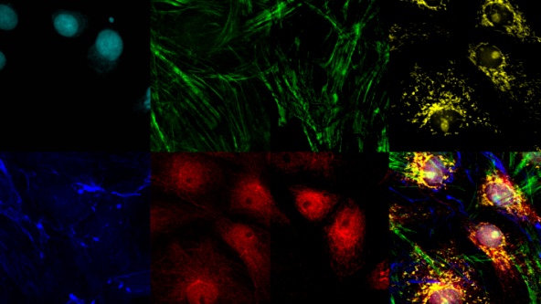
라이브 셀 초고해상도 이미징: 작은 대상을 큰 그림으로
Experts


이 웨비나에서 Olympus 이미징 전문가 Stefan, Lauren, Chunsong은 초고해상도 이미지를 더 간편하게 생성하는 방법에 대해 이야기합니다.
Digital Image Processing: Point and Local Operation Filters
Experts


Images captured with a light microscope are never true representations of the specimen; there are always sources of error that must be controlled. In this webinar, we will discuss how these sources of error can be managed.
Think Deep, See Deeper with Near-Infrared Laser Scanning Confocal Microscope
Experts

If you’re interested in having more fluorescent markers in your sample, imaging deeper into your sample at high resolution, and conducting live cell imaging with minimal phototoxicity, check out this on-demand webinar from Olympus imaging experts.
Light Sheet Microscopy for Deeper Insight into Life
Experts

In this webinar, you’ll learn how the Alpha3 light sheet microscope combines very thin optical sectioning and high-quality Olympus optics for high-resolution 3D imaging of both live and fixed biological samples.
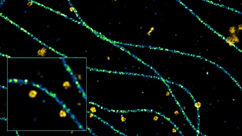
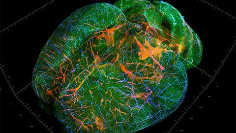
나노미터 크기의 여러 세포 내 구조를 3D로 이미징하기
Experts

Abbelight의 공동 창업자인 Nicolas Bourg 박사가 샘플의 안정적이고 뚜렷한 고품질 단분자 이미지를 15nm 해상도에서 얼마나 쉽고 빠르게 3D로 획득할 수 있는지 이야기합니다.
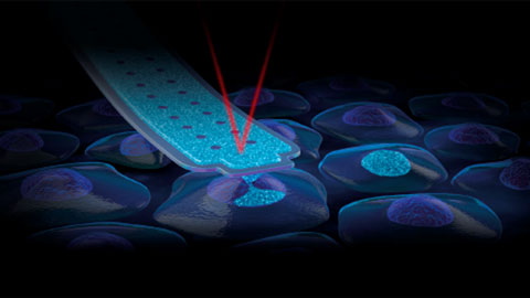
FluidFM: 핵 내 직접 전달을 통한 새로운 CRISPR 유전자 편집 방식
Experts
이 웨비나에서는 Cytosurge의 Paul Monnier가 유전자 편집의 가장 큰 과제 중 하나, 즉 유전 물질을 핵으로 전달하는 어려운 과제를 해결하는 방법을 보여드립니다.Paul은 핵막을 부드럽게 관통하여 시약을 핵으로 직접 전달할 수 있는 FluidFM 나노 주사기에 대해 이야기하겠습니다.
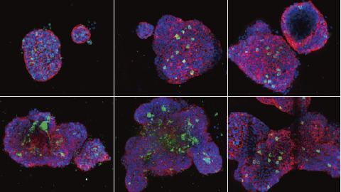
3D 분석: 지능형 소프트웨어, 통찰력 있는 분석
Experts


3D 분석: 지능형 소프트웨어, 통찰력 있는 분석 이 웨비나에서 스페로이드 및 마이크로플레이트 기반 분석에 대한 최적의 통계 데이터를 얻는 방법에 대해 논의합니다. 이러한 데이터를 사용하면 화합물에 대한 3D 모형의 반응을 정량화하고 다양한 농도와 같은 다른 처치의 효과를 쉽게 비교할 수 있습니다.
Multiplexing and Deep Tissue Imaging with Near-Infrared Confocal Laser Scanning Microscopy
Experts


In this webinar, you’ll learn about fluorescence multiplexing and deep tissue imaging using near-infrared (NIR) laser light.

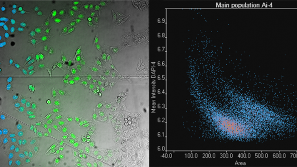
최신 슬라이드 스캐닝: 고정 샘플에서의 단세포 표현형
Experts


4월에는 지구의 날과 2021 올해의 이미지상 수상자를 기념하였으며, 여러분께서는 다양한 이미지를 감상하셨습니다. 그중 최고 인기 이미지를 여기에서 확인하세요.