Evident LogoOlympus Logo

Ask the Experts

The Use of Multiplexing in Microscopy for Better Understanding the Skin Immune System in the Context of the Tissue

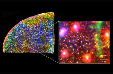
The skin is the first line of defense and the immune system’s biggest barrier for combating pathogens. Being able to accurately characterize and identify immune cell subtypes, tissues structures, and cell distribution in the skin under steady-state conditions provides a powerful tool for understanding the first immunological strategies and biological processes that occur in the presence of pathogens. In this webinar we will review technical aspects involved in the experimental process and explore how complementary imaging technologies might assist us to better understand the immune system.

The presentation is divided into three parts. First, an introduction of the Hugh Green Cytometry Centre will be presented and an overview of the histology and bioimaging technological platforms available. Second, the multiplexing methodology will be discussed, where several topics need to be considered for the design and development of a successful polychromatic panel for microscopy. Finally, preliminary results from a research project will be presented that constitutes part of a diploma program from The Royal Microscopical Society. The project focuses on the identification of immune cell types in the whole mount skin in relation to tissues structures (e.g., blood vessels and lymphatic network). It also centers on the immune cells’ distribution in the tissue as a first barrier of defense against pathogens.

Presenter: Alfonso J. Schmidt
Senior Staff Scientist, Malaghan Institute of Medical Research

Alfonso has a decade of experience working in a Shared Resource Lab (SRL) with a vast knowledge in histology, fluorescent microscopy, and image analysis. His work has been focused in maximizing the capabilities of the equipment available and in creating technical protocols and training modules for the scientific community. Currently, Alfonso oversees the Histology and Bioimaging Facility as part of the Hugh Green Cytometry Centre (HGCC) at the Malaghan Institute of Medical Research. Wellington, New Zealand.  

관련 제품

컨포칼 레이저 스캐닝 현미경

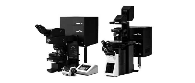
FV3000

  • 검류계 전용(FV3000) 또는 검류계/공진(FV3000RS) 하이브리드 스캐너 구성에 사용 가능
  • 모든 채널에 정확한 고효율 TruSpectral 검출
  • 고광도 및 저광독성으로 라이브 세포 이미징 최적화
리서치 슬라이드 스캐너

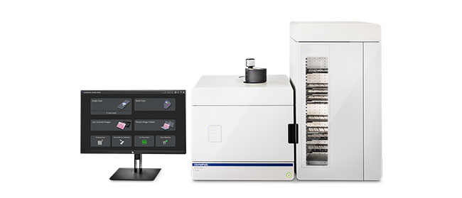
VS200

VS200 연구 슬라이드 스캐너는 뇌, 암, 줄기 세포 및 신약 개발 연구를 비롯하여 대량의 샘플에 대한 정량 분석이 필요한 응용 부문의 연구 속도와 효율을 향상합니다. 고해상도 및 고품질 슬라이드 이미지를 쉽고 빠르게 확보하고 데이터를 쉽게 분석, 공유 및 기록할 수 있습니다. VS200 시스템은 명시야, 형광, 암시야, 위상차 및 단순 편광이라는 다섯 가지 이미지화 모드를 제공합니다. 자동 스캐닝 시 한 번에 최대 210개의 슬라이드를 로드할 수 있습니다.

  • 2X에서 100X까지 고해상도 전체 슬라이드 이미지화
  • 다양한 크기의 슬라이드 및 관찰 방법 수용
  • 명시야에서 형광 다중화에 이르는 간단하고 강력한 워크플로
  • 수동 명시야 스캐닝에서 AI 인식 및 완전 자동 스캐닝에 이르기까지 소프트웨어 및 하드웨어에서 고유하게 구성 가능

The Use of Multiplexing in Microscopy for Better Understanding the Skin Immune System in the Context of the Tissue

The skin is the first line of defense and the immune system’s biggest barrier for combating pathogens. Being able to accurately characterize and identify immune cell subtypes, tissues structures, and cell distribution in the skin under steady-state conditions provides a powerful tool for understanding the first immunological strategies and biological processes that occur in the presence of pathogens. In this webinar we will review technical aspects involved in the experimental process and explore how complementary imaging technologies might assist us to better understand the immune system.

The presentation is divided into three parts. First, an introduction of the Hugh Green Cytometry Centre will be presented and an overview of the histology and bioimaging technological platforms available. Second, the multiplexing methodology will be discussed, where several topics need to be considered for the design and development of a successful polychromatic panel for microscopy. Finally, preliminary results from a research project will be presented that constitutes part of a diploma program from The Royal Microscopical Society. The project focuses on the identification of immune cell types in the whole mount skin in relation to tissues structures (e.g., blood vessels and lymphatic network). It also centers on the immune cells’ distribution in the tissue as a first barrier of defense against pathogens.

Presenter: Alfonso J. Schmidt
Senior Staff Scientist, Malaghan Institute of Medical Research

Alfonso has a decade of experience working in a Shared Resource Lab (SRL) with a vast knowledge in histology, fluorescent microscopy, and image analysis. His work has been focused in maximizing the capabilities of the equipment available and in creating technical protocols and training modules for the scientific community. Currently, Alfonso oversees the Histology and Bioimaging Facility as part of the Hugh Green Cytometry Centre (HGCC) at the Malaghan Institute of Medical Research. Wellington, New Zealand.  

관련 제품

컨포칼 레이저 스캐닝 현미경

FV3000

  • 검류계 전용(FV3000) 또는 검류계/공진(FV3000RS) 하이브리드 스캐너 구성에 사용 가능
  • 모든 채널에 정확한 고효율 TruSpectral 검출
  • 고광도 및 저광독성으로 라이브 세포 이미징 최적화
리서치 슬라이드 스캐너

VS200

VS200 연구 슬라이드 스캐너는 뇌, 암, 줄기 세포 및 신약 개발 연구를 비롯하여 대량의 샘플에 대한 정량 분석이 필요한 응용 부문의 연구 속도와 효율을 향상합니다. 고해상도 및 고품질 슬라이드 이미지를 쉽고 빠르게 확보하고 데이터를 쉽게 분석, 공유 및 기록할 수 있습니다. VS200 시스템은 명시야, 형광, 암시야, 위상차 및 단순 편광이라는 다섯 가지 이미지화 모드를 제공합니다. 자동 스캐닝 시 한 번에 최대 210개의 슬라이드를 로드할 수 있습니다.

  • 2X에서 100X까지 고해상도 전체 슬라이드 이미지화
  • 다양한 크기의 슬라이드 및 관찰 방법 수용
  • 명시야에서 형광 다중화에 이르는 간단하고 강력한 워크플로
  • 수동 명시야 스캐닝에서 AI 인식 및 완전 자동 스캐닝에 이르기까지 소프트웨어 및 하드웨어에서 고유하게 구성 가능
Experts
Alfonso J. Schmidt
Senior Staff Scientist
Malaghan Institute of Medical Research

Alfonso has a decade of experience working in a shared resource lab (SRL) with a vast knowledge in histology, fluorescent microscopy, and image analysis. His work has been focused in maximizing the capabilities of the equipment available and in creating technical protocols and training modules for the scientific community. Currently, Alfonso oversees the Histology and Bioimaging Facility as part of the Hugh Green Cytometry Centre (HGCC) at the Malaghan Institute of Medical Research in Wellington, New Zealand.

The Use of Multiplexing in Microscopy for Better Understanding the Skin Immune System in the Context of the Tissue2024년12월13일
죄송합니다. 이 페이지는 해당 국가에서 사용할 수 없습니다.
Ask the Expert Sign-up

By clicking subscribe you are agreeing to our privacy policy which can be found here.

Sorry, this page is not
available in your country.

죄송합니다. 이 페이지는 해당 국가에서 사용할 수 없습니다.