
While our recently announced 2020 Image of the Year Competition winners justifiably took the top spots this month, we wanted to showcase the other images that caught people’s eye in our monthly round up.
Our theme for this month is the letter A for April, algae, and ants.
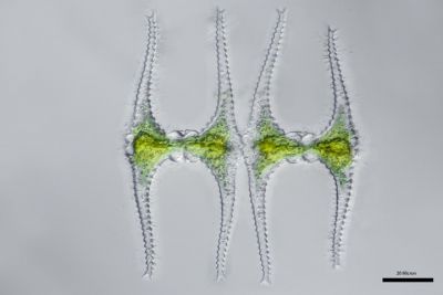
This one kept some people guessing, but many correctly identified this image as a genus of green algae called Staurastrum. We particularly enjoyed that a couple people suggested that it might be a Star Wars TIE fighter. Perhaps we unknowingly found George Lucas’ inspiration for the ship’s design!
Image courtesy of Linden Gledhill.
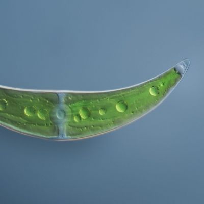
This month’s image from Håkan Kvarnström’s series, "Exploring the Microscopic World with X Line Objectives," is not a very small plantain, but rather a green alga called Closterium. The genus Closterium—living exclusively in freshwater environments—is one of the closest single-cell relatives to land plants and dates back 450 million years.
Håkan explains that up until recently, it had been assumed that the alga evolved the capability to source essential nutrients for its survival after it arrived on land by forming a close association with beneficial fungi. However, recent research shows the algal ancestor living in the Earth's waters already possessed the set of genes needed to detect and interact with the beneficial fungi and the ability to live on land, making it possible for the alga to survive out of the water and to colonize the Earth.
Without the development of this pre-adapted capability in alga, the Earth could be a very different place today. However, the origin of these genes and their potential correlation with land colonization remain a mystery.
Description and image courtesy of Håkan Kvarnström. Captured by focus stacking images taken with DIC (40X) and Olympus X Line objectives.
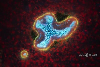
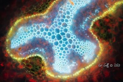
Photographed in ultraviolet-induced excitation microscopy, these images show two captures of a 30-micron section through the petiole of the Harts Tongue Fern, commonly found in Europe and the eastern US.
Images courtesy of Karl Gaff.
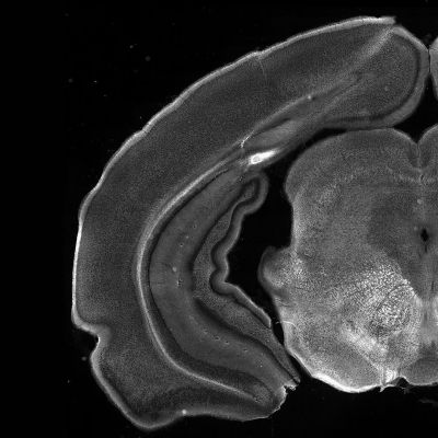
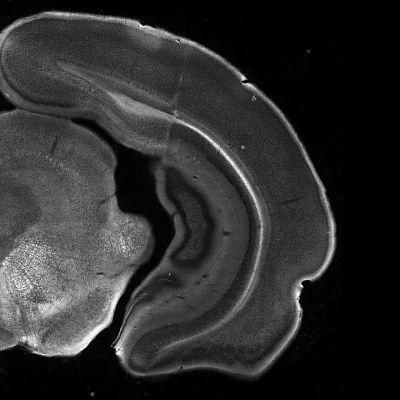
Did you know? A mouse’s brain contains about 0.07% as many neurons as you’d find in an average human! This image shows the whole coronal section of the brain.
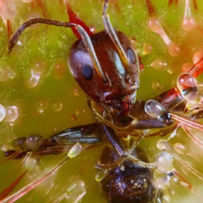
This image shows an ant on a deathbed of sundew, a carnivorous plant that traps prey in the sticky hairs located on the leaves!
Image courtesy of Ralph Grimm.
Bonus video!
This month Benedikt Pleyer captured our attention with his Spring Flowers of the Microworld.
“As soon as the icy grip of winter subsides, life pushes back into the emptiness this season leaves behind. Just think of flowers like snowdrops growing effortlessly through the thawing snow.
The microworld has its own spring pioneers: diatoms! These glass creatures proliferate explosively in the cool and silicon-rich water coming down the flanks of mountains where the snowmelt has set in with all its might.
Surirella, shown here in the footage, is my personal favorite. These diatoms start to form a 2 cm deep, golden brown layer at the bottom of mountain springs and emanate the silent hum of Vivaldi’s Four Seasons. Surrrrirella!”
Video and caption courtesy of Benedikt Pleyer. Captured at 200x-400x magnification and sped up 30-60 minutes using DIC. Captured on an Olympus BX53 microscope.
To see more images like these, be sure to follow us on Instagram at @olympuslifescience!
Interested in sharing your own images? Visit our image submission site.
Related Content
Scientific Works of Art: Announcing the Winners of Our 2020 Global IOTY Award
Feline Fine in Springtime: Our Most Popular Microscope Images for March 2021
Connecting with Curiosity: A Conversation with Dr. Gabrielle Corradino
