세포 주기 표지자 Fucci를 사용한 줄기 세포 생리 상태의 시각화 (Visualization of Physiological State of Stem Cells Using Cell Cycle Marker Fucci)
|
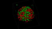
Fucci로 세포 주기가 표지된 CD133-양성 인간 위암세포는 타원체를 포함하고 있으며, 배양하여 장시간 연속적으로 관찰할 수 있습니다. (Spheroids consisted of CD133-positive human gastric cancer cells
with cell cycle marker Fucci were cultured and observed continuously over an extended period.)슬라이드의 상부 패널은 타원체를 배양액 없이 배양함으로써 세포 주기를 멈춰 줄기세포 상태를 유지합니다.한편,
하단 패널에는 타원체를 세포 분열에 필요한 혈청과 함께 배양하였습니다.이 이미지는 타원체들이 단층 배양 상태로 전환하는 모습을
나타냅니다. (줄기세포와 유사한 상태를 상실)컨포컬 레이저 스캐닝 현미경(Confocal Laser Scanning Microscope) FV10i로 두꺼운 타원체를 입체적으로 관찰할 수 있습니다.
*이 이미지는 컨포컬 레이저 스캐닝 현미경(Confocal Laser Scanning Microscope) FV10i로 48시간 동안 연속으로 촬영하였습니다. |
이미지 데이터 제공 :
Shuya Yano, Toshiyoshi Fujiwara
Department of Gastroenterological Surgery Transplant and Surgical oncology, Okayama University Graduate School of Medicine, Dentistry and Pharmaceutical Sciences.
Reference:
Yano S, Tazawa H, Hashimoto Y, Shirakawa Y, Kuroda S, Nishizaki M, Kishimoto H, Uno F, Nagasaka T, Urata Y, Kagawa S, Hoffman RM, Fujiwara T. 유전자 재조합 종양 용해성 아데노 바이러스(genetically engineered oncolytic adenovirus)는 정지 암 줄기세포와 같은 세포(quiescent cancer stem-like cells)를 S/G2/M 단계로 유도하고 가둡니다.Clin Cancer Res. December 1, 2013 19:6495-6505.
잠재 암의 Telomelysin (OBP-301)에 의한 줄기세포의 활성화 및 세포 파괴 (Activation and Cell Destruction of Stem Cells from Sleeping Cancer by Telomelysin (OBP-301))
|
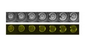
CD133-양성, 줄기세포 상태 위암 세포와 타원체와 배양한 세포주기 표지자 Fucci를 telomelysin (OBP-301), cisplatin 혹은 방사선으로 처리합니다. (The CD133-positive, stem-like human gastric cancer cells with the cell cycle marker Fucci which had been
cultured as spheroids were treated with either telomelysin (OBP-301), cisplatin or radiation.)Cisplatin 혹은 방사선으로 처리한 암세포 덩어리는 같은 크기로 유지되며, 세포 대부분은 세포주기 G1기를 넘기지 않습니다.
(빨간색 참조) (The clumps of cancer cells treated with cisplatin or radiation remained the same size and the majority of the cells did not proceed the cell cycle beyond the G1 phase (seen in red).)한편, telomelysin으로 처리한 암세포 덩어리는 빨강에서 노랑 및 초록으로 색이
변하며, 크기는 점점 작아집니다.이는 세포줄기와 같은 세포 상태로 세포 주기를 정지하는 p53과 p21의 발현을 telomelysin이 억제하는 것을 의미합니다. (This indicates that
telomelysin inhibits the expression of p53 and p21, both of which are responsible for arresting the cell cycle of stem-like cells.)Telomelysin은 E2F-1의 발현을 증가시켜서, 세포 주기가 정반대로 활성화됩니다. (Telomelysin also increases the expression of E2F-1, which leads to the activation of the cell cycle by contraries.)
*이 이미지는 컨포컬 레이저 스캐닝 현미경(Confocal Laser Scanning Microscope) FV10i로 8일 동안 연속으로 촬영하였습니다. |
이미지 데이터 제공 :
Shuya Yano, Toshiyoshi Fujiwara
Department of Gastroenterological Surgery Transplant and Surgical oncology, Okayama University Graduate School of Medicine, Dentistry and Pharmaceutical Sciences.
Reference:
Yano S, Tazawa H, Hashimoto Y, Shirakawa Y, Kuroda S, Nishizaki M, Kishimoto H, Uno F, Nagasaka T, Urata Y, Kagawa S, Hoffman RM, Fujiwara T. 유전자 재조합 종양 용해성 아데노 바이러스(genetically engineered oncolytic adenovirus)는 정지 암 줄기세포와 같은 세포(quiescent cancer stem-like cells)를 S/G2/M 단계로 유도하고 가둡니다. Clin Cancer Res. December 1, 2013 19:6495-6505.
Fucci Induced Spheroid of HT29 Cell Line
이미지 데이터 제공 :
Dr. Yuji Mishima, Dr. Kiyohiko Hatake
Clinical Chemotherapy, Cancer Chemotherapy Center, Japanese Foundation for Cancer Research
전이 생태 및 다층 근육 아세포 병풍구조 내의 섬유아세포의 세포-세포 연결
|
이미지 데이터 제공 :
Eiji Nagamori, Ph.D Masahiro Kino-oka, Ph.D. Department of Biotechnology, Graduate School of Engineering, Osaka University |
YFP를 전사인자로 사용한 제프라피쉬 배아에서 레티노산 신호의 시각화 (Visualizing Retinoic Acid Signaling in A Zebrafish Embryo Using YFP as a Reporter)
이미지 데이터 제공 :
Satoshi Shimozono, Ph.D. Atsushi Miyawaki, M.D., Ph.D.
Laboratory for Cell Function Dynamics, Advanced Technology Development Core, RIKEN Brain Science Institute
Reference:
Shimozono S. et al. 발달 중인 배아의 점진적인 내인성 레티노산의 시각화. (Visualization of an endogenous retinoic acid gradient across embryonic development.)Nature 496, 363-366 (18 April 2013)
항체 의존성 세포 독성(Antibody Dependent Cellular Cytotoxicity, ADCC)의 관찰
|
RPMI 4788 세포(인간 결장암 세포주)는 항체의약품 cetuximab으로 처리하고 NK 세포(Natural Killer Cell)와 함께 배양하면 ADCC는 NK 세포 추가 후에 FV10i를 사용하여 관찰할 수 있습니다.
(RPMI 4788 cells (human colon cancer cell line) were treated with an antibody drug, cetuximab, and co-cultured with natural killer (NK) cells ADCC was observed using the FV10i after addition of NK cells)
Cetuximab: Alexa Fluor 647 (red) NK cells: ZsGreen (green) 죽은 세포 감지: DAPI (blue) |
이미지 데이터 제공 :
Dr. Yuji Mishima, Dr. Kiyohiko Hatake
Clinical Chemotherapy Department, The Cancer Chemotherapy Center of the Japanese Foundation for Cancer Research
다양한 농도의 항암제 시스플라틴(Cisplatin)의 효과 관찰
| 항암제 시스플라틴(Cisplatin) 처리한 HT-29 세포의 세포주기는 HT-29를 발현하는 Fucci를 저속 촬영(Time-lapse)하여 관찰하였습니다.세포는 대조군( 0 ug/ml), 저농도(0.25 ug/ml), 고농도(2.5 ug/ml)의 서로 다른 농도의 Cisplatin으로 처리하여 35mm 유리 바닥 디쉬에서 48시간 동안 배양하였습니다. |
이미지 데이터 제공 :
Dr. Yuji Mishima, Dr. Kiyohiko Hatake
Clinical Chemotherapy Department, The Cancer Chemotherapy Center of the Japanese Foundation for Cancer Research
긴 돌기 세포-세포 융합(Cell–cell Fusion with Long Projections) 동안 포스포이노시티드(Phosphoinositides)의 Localization
| 1 µg/ml 독시사이클린(doxycycline)에서 생성된 RAW264.7 세포는 Reporter를 발현하며, 이를 10 ng/ml RANKL로 48시간 동안 자극합니다. (RAW264.7 cells expressing the reporter constructs in the presence of 1 µg/ml doxycycline were stimulated with 10 ng/ml RANKL for 48 h.)각 Localization이 PtdIns(4,5)P2은 빨간색으로 보이며, PtdIns(3,4,5)P3은 녹색으로 보입니다.프레임은 총 34분동안 2분마다 25µm 간격으로 찍습니다. |
이미지 데이터 제공 :
Tsukasa Oikawa, Ph.D.
Department of Molecular Biology, Hokkaido University Graduate School of Medicine
Reference:
Oikawa T, et al. 원기둥 모양의 podosomes/invadopodia의 Tks5-의존 형성은 세포-세포 융합에 영향을 줍니다.J Cell Biol. 197(4):553-568(2012).
헬라 세포(HeLa Cell)*1
|
염색 시약: YFP (Actin)
렌즈: 60XW NA1.2 레이저 파장: 473nm 시간 간격: 매 3.5분 (총 12시간) |
쥐의 뇌
| 쥐의 뇌를 DAPI(nuclei - blue), Alexa 488(Actin - green), Alexa 568(Neurofilament - red)로 형광 표지합니다.9개의 관심 영역을 1024x1024로 Z축 14개 부분 이상으로 자동 획득하고, FV10i 소프트웨어로 이미지를 이어 붙입니다. |
헬라 세포(HeLa Cell)*1
|
시간 간격: 밤새 매 20분 간격으로 촬영
Green: GFP Magenta: Mito Tracker Red |
*1 비록 헬라 세포가 의료 연구에서 가장 중요한 세포주가 되었다고 해도, 과학에 대한 Henrietta Lacks의 공헌이 동의를 받지 않은 것이었다는 것을 인정해야만 합니다. 이로 인해 면역학, 전염병, 암에 대한 중요한 발견이 이루어졌지만 사생활, 윤리, 의학적 동의에 대한 중요한 논의도 촉발되었습니다.
Henrietta Lacks의 삶과 현대 의학에 대한 그녀의 공헌을 알아보려면 여기를 클릭하세요.
http://henriettalacksfoundation.org/