EVIDENT Image of the Year Award 2022We're thrilled to announced the winners of the Evident Image of the Year Award 2022. We thank everyone who entered, and we look forward to your participation again next year. |
|

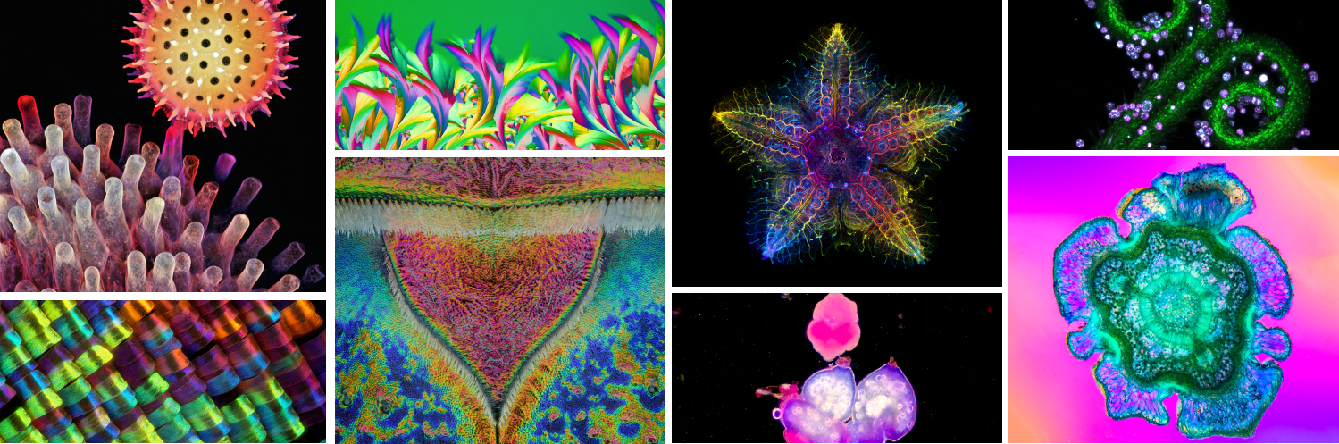
The Global WinnerThe global winning image was taken by Laurent Formery (USA). Nervous system of a juvenile sea star (Patiria miniata) about 1 cm wide. Labeled with an antibody against acetylated tubulin after optical clearing, and captured using a color-coded Z-projection. | 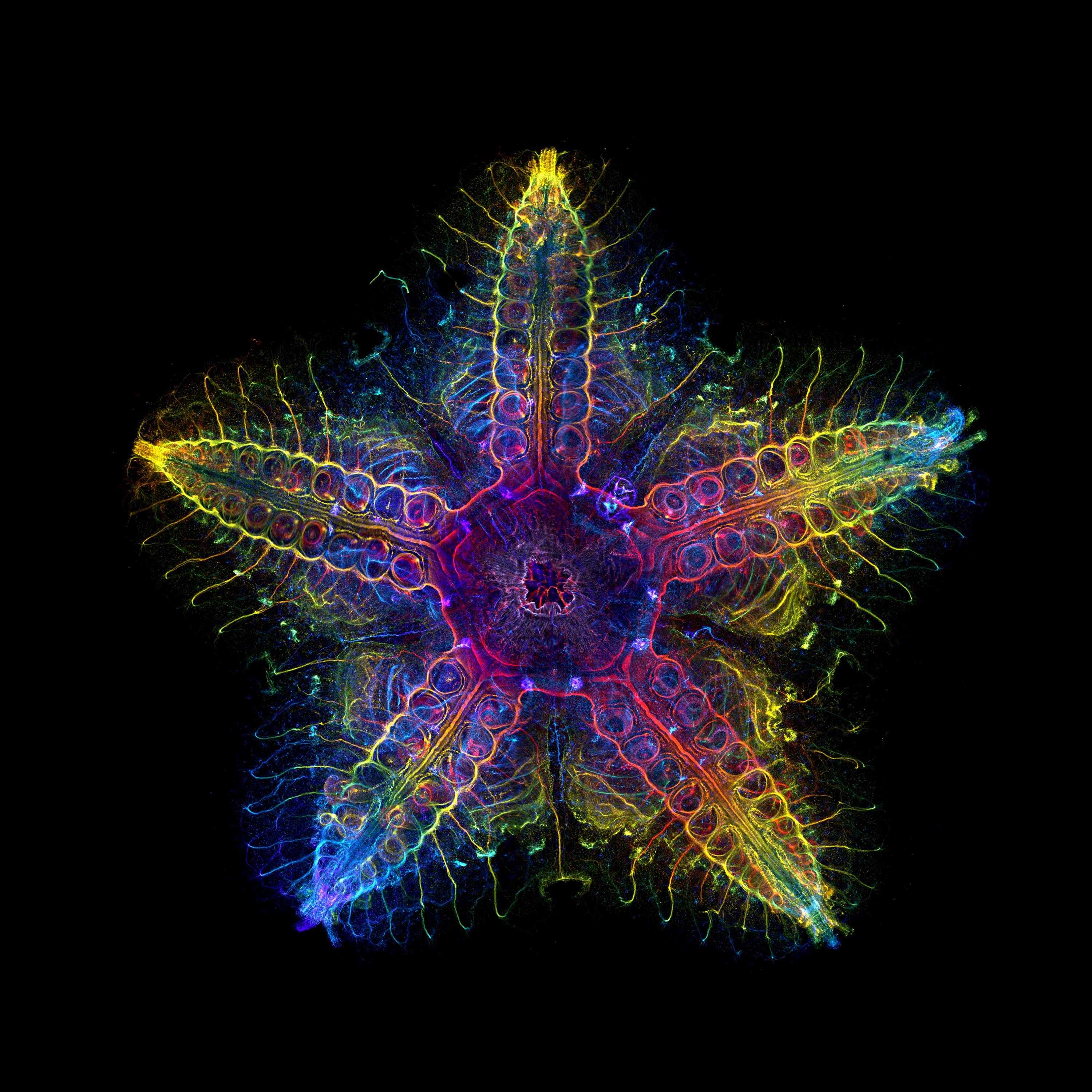 |
|---|
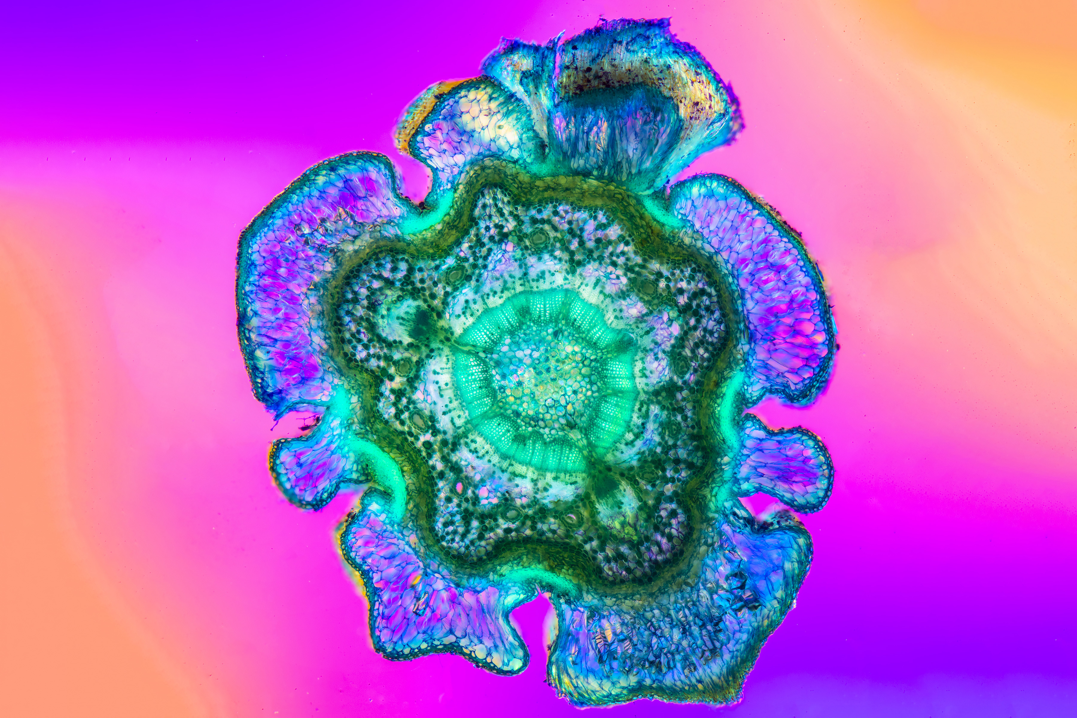
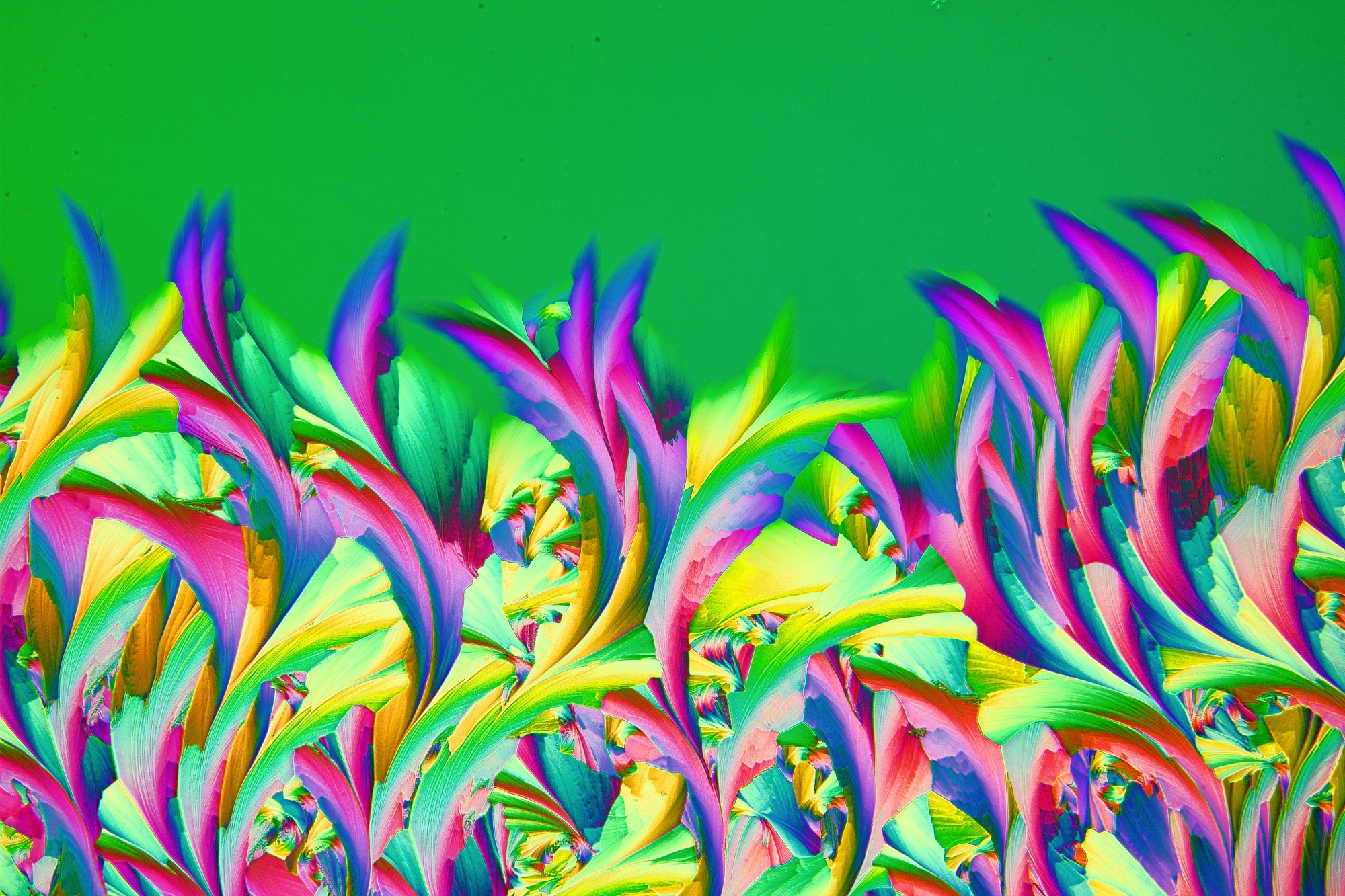 | The Materials Science WinnerThe winning image for our materials science category was taken by Shyam Rathod (India). Crystal of a topical medicine for wart treatment named ABE, which is available in Poland. The dew was blown on a microscope slide with a straw and was captured in a single frame. Retarder and a two-cross polarization filter were used to bring out the colors. |
|---|
Regional Winners
Americas The winning image for the Americas was captured by Igor Siwanowicz (USA). Depth color-coded projection showing a germinating pollen grain of a morning glory attached to the stigma. | Europe, the Middle East, and Africa The winning image for EMEA was captured by Javier Ruperez (Spain). Scales of the wing of the Urania rhipheus moth. Captured at 20X. | Asia-Pacific The winning image for Asia was taken by Jiao Li (China). Edelweiss stamens, which were scanned and reconstructed in three dimensions using laser scanning confocal microscopy. |
Honorable Mentions
 Lateral head of a zebrafish, 72 hours after fertilization. | 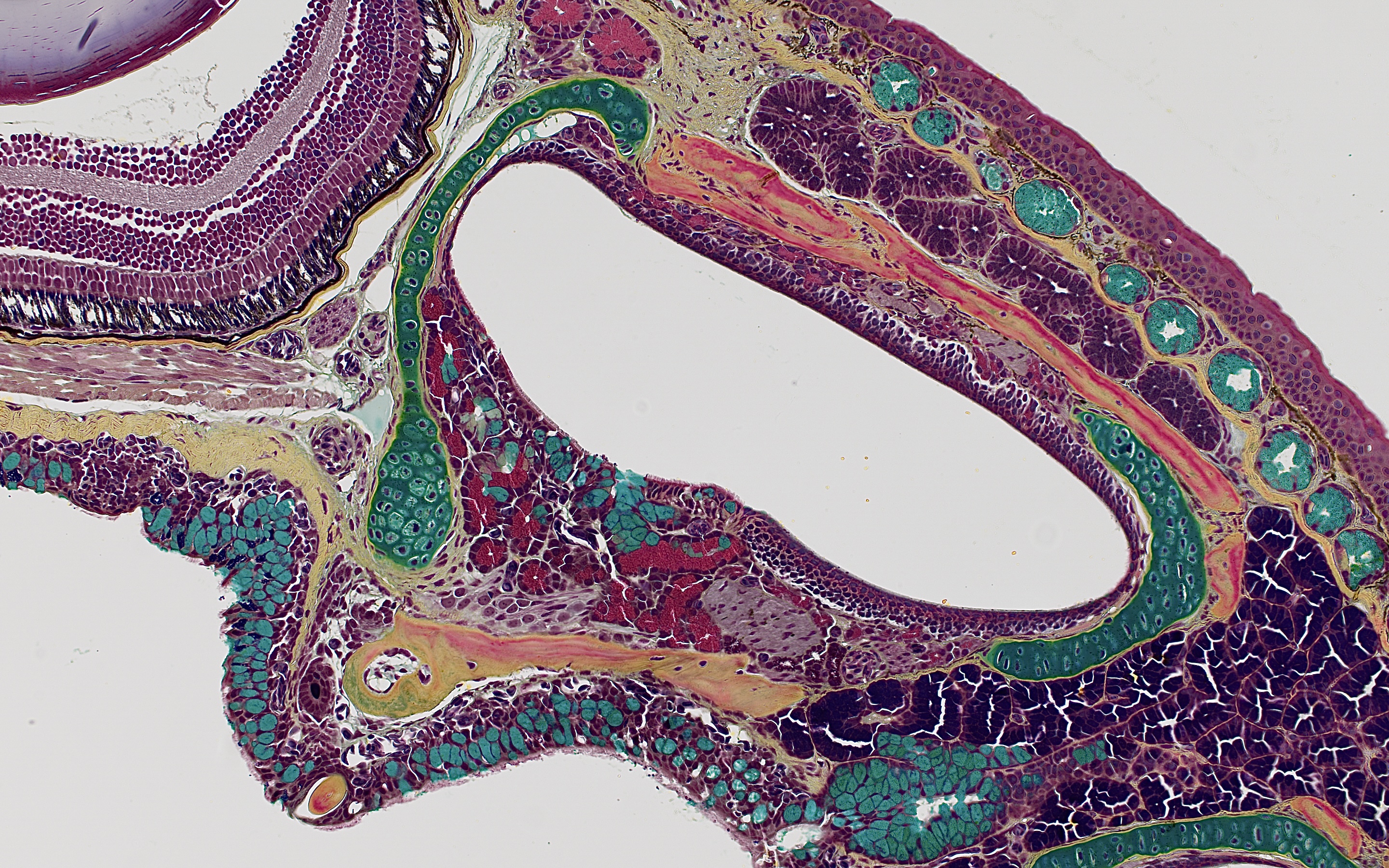 Dorsal view of a red-backed salamander skull. Stained with Movat's pentachrome technique. | 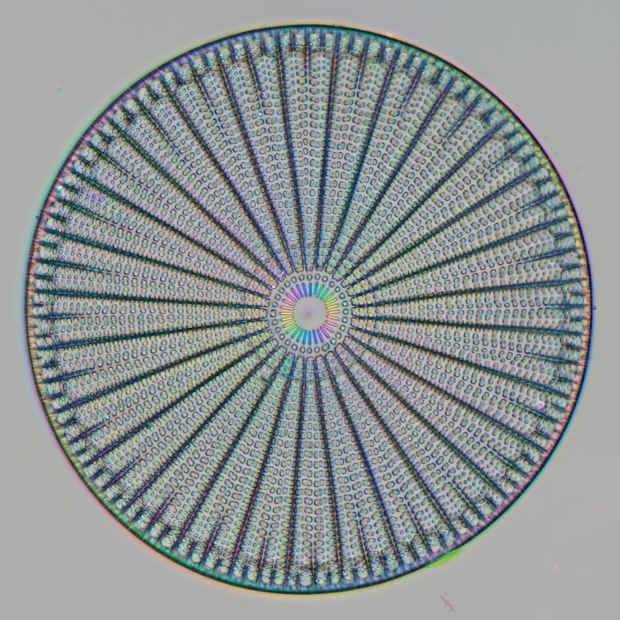 Diatoms are unicellular organisms, which can be found in the oceans, waterways, and soils of the world. | 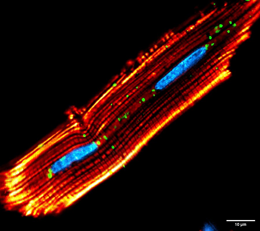 Trichromatic confocal imaging of isolated cardiomyocytes from adult rats, showing the nucleus, lysosomes, and mitochondria, respectively. | 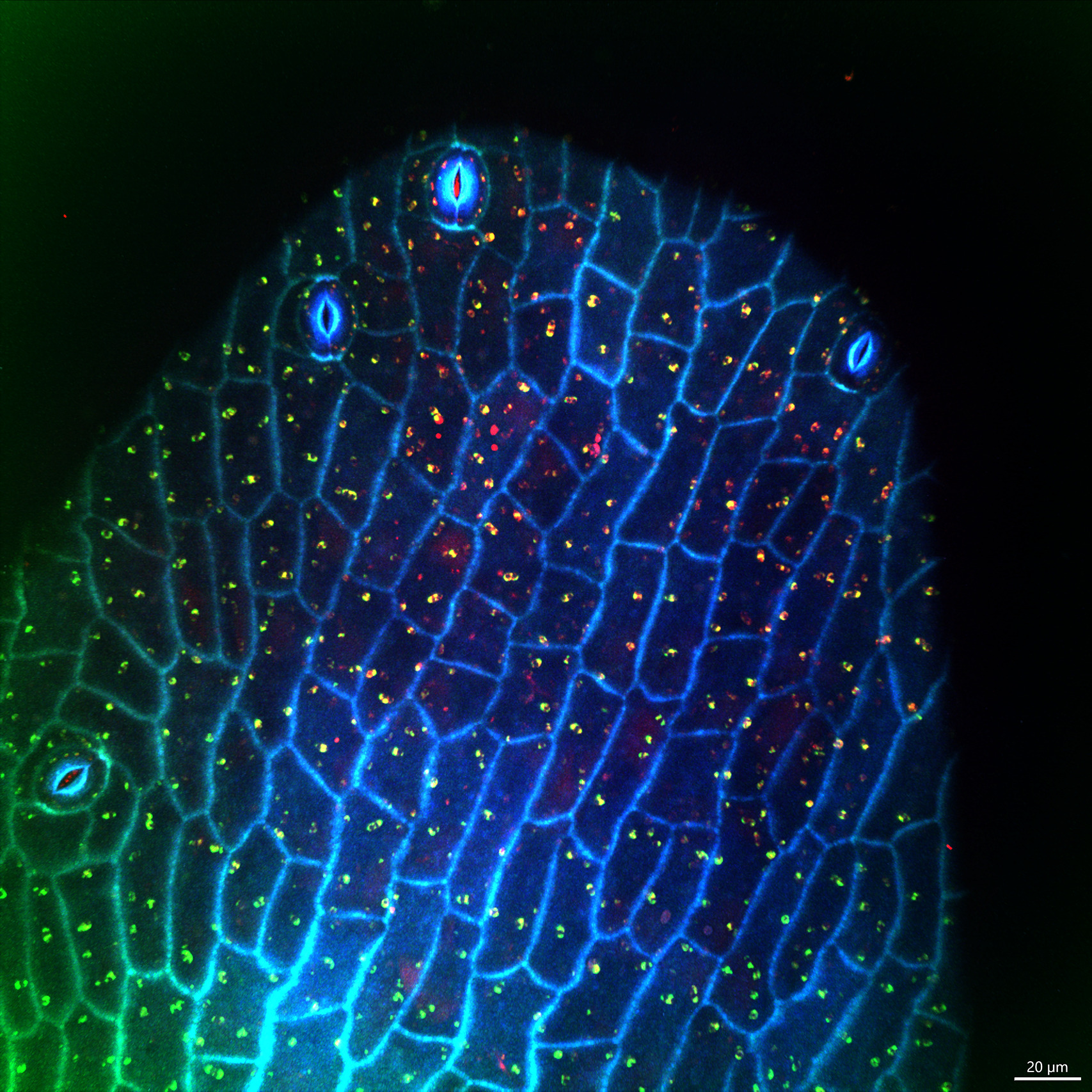 Autofluorescence of the tip of the stamens of flowers at 405 nm, 488 nm, and 561 nm, emitting a greenish blue and emerald green color like peacock feathers. |
 Cross section of a blue spruce (Picea pungens) branch. The image is a panorama of several photos that were focus stacked and captured with a 5X objective. | 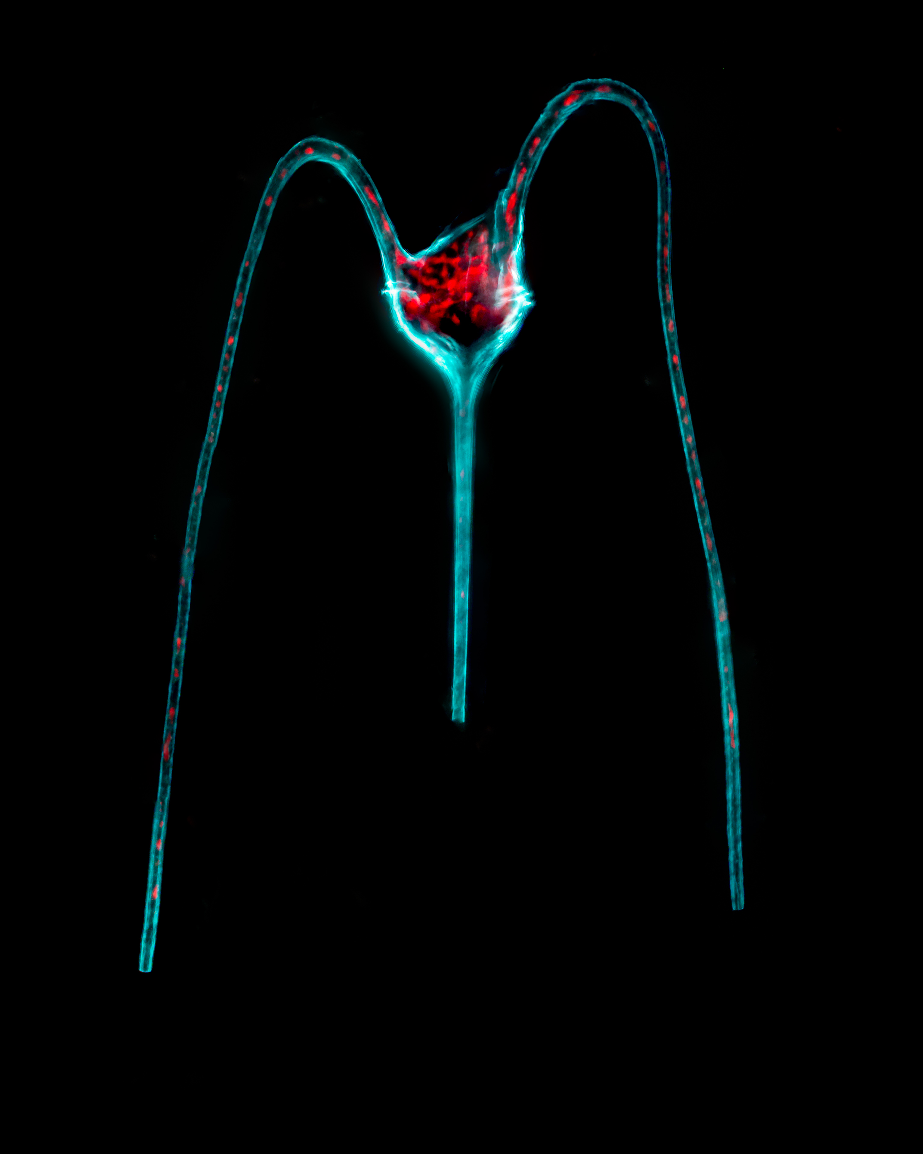 Tripos macroceros. A unicellular microalga with three horns. The antapical horns are bent toward the single apical horn. The chlorophyll is displayed in red, where the chloroplasts sit. | 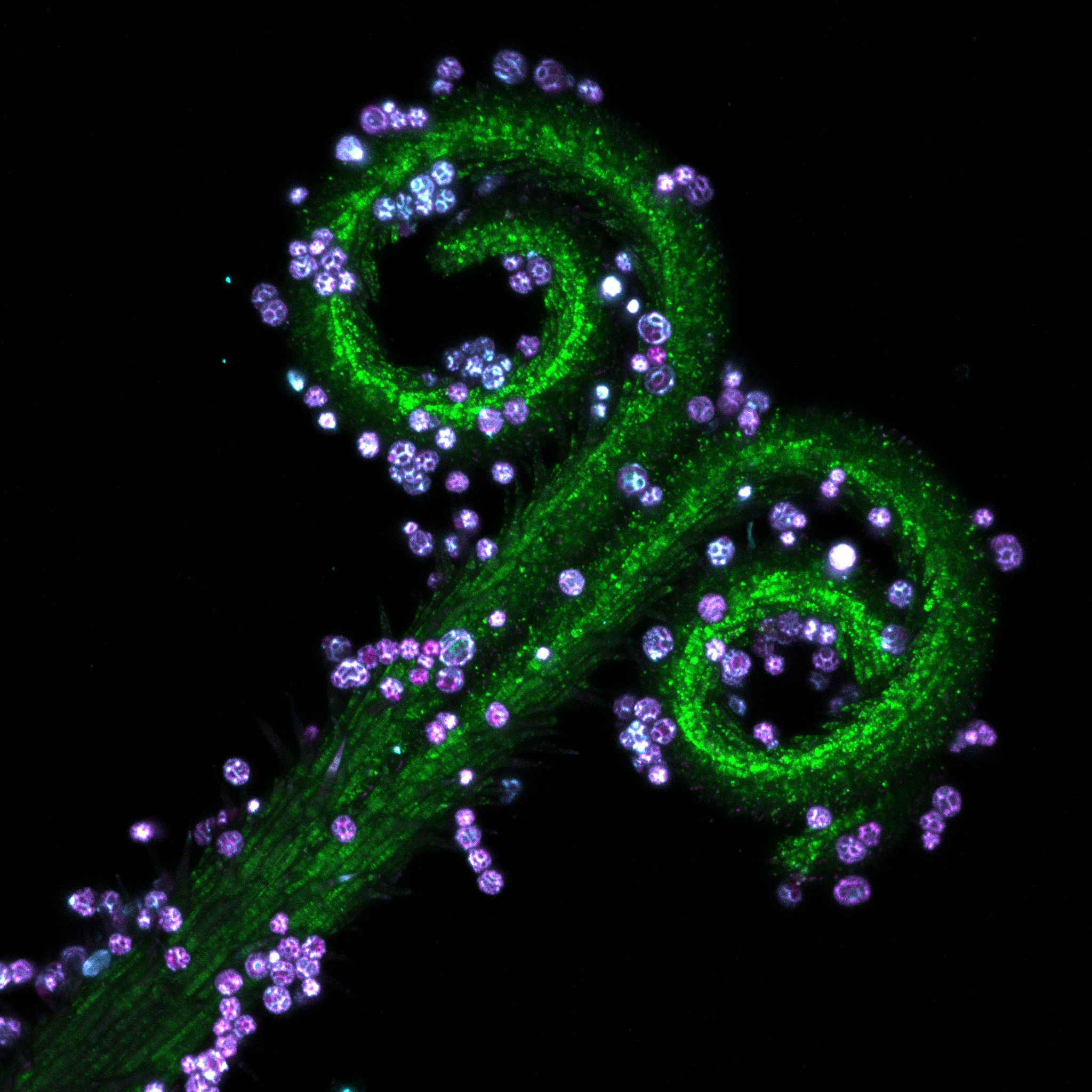 Pistils of dandelion were collected and made into slide samples. Different fluorescence patterns of its parts were observed using confocal microscopy. | 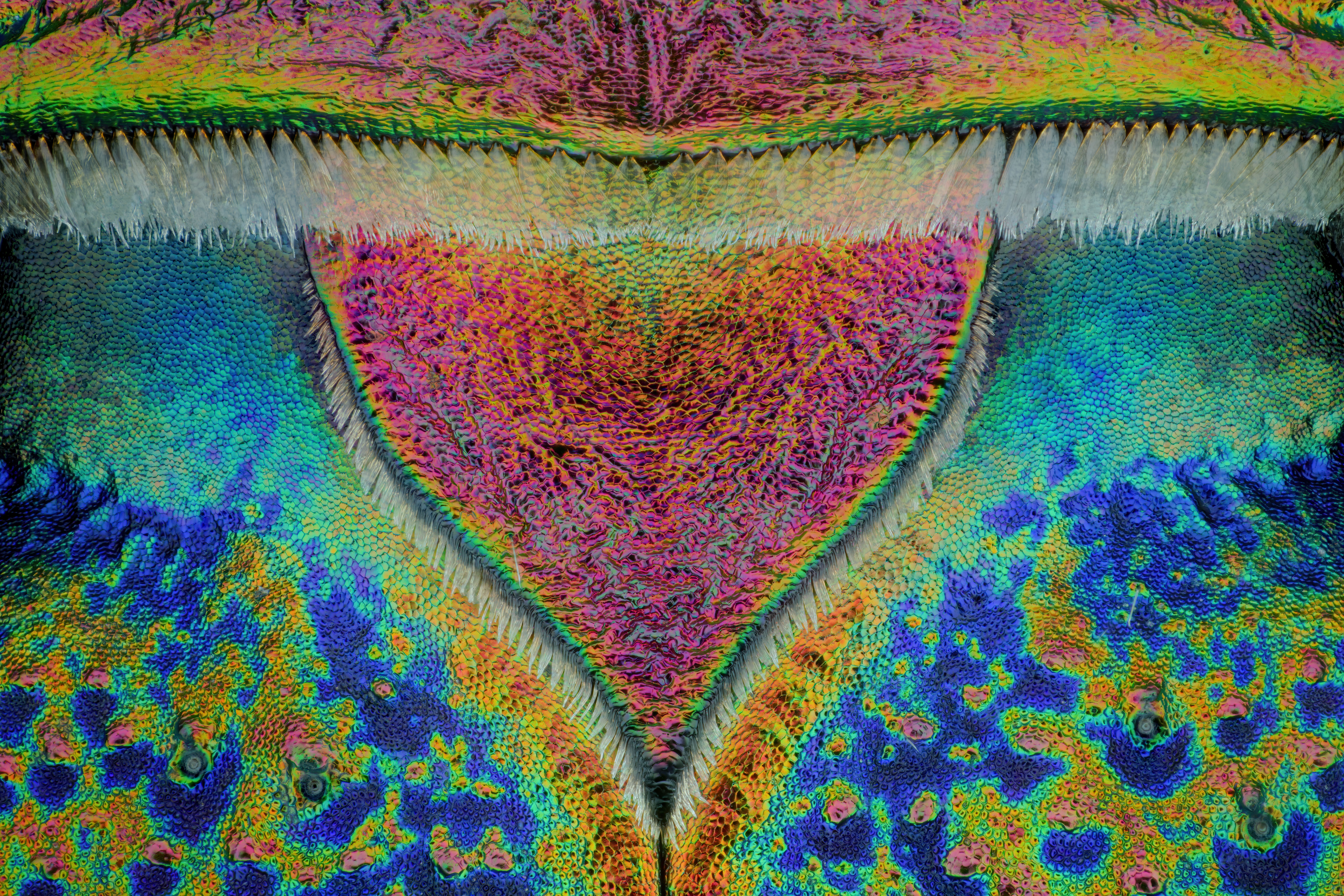 Scutellum of a tiger beetle 20:1 | 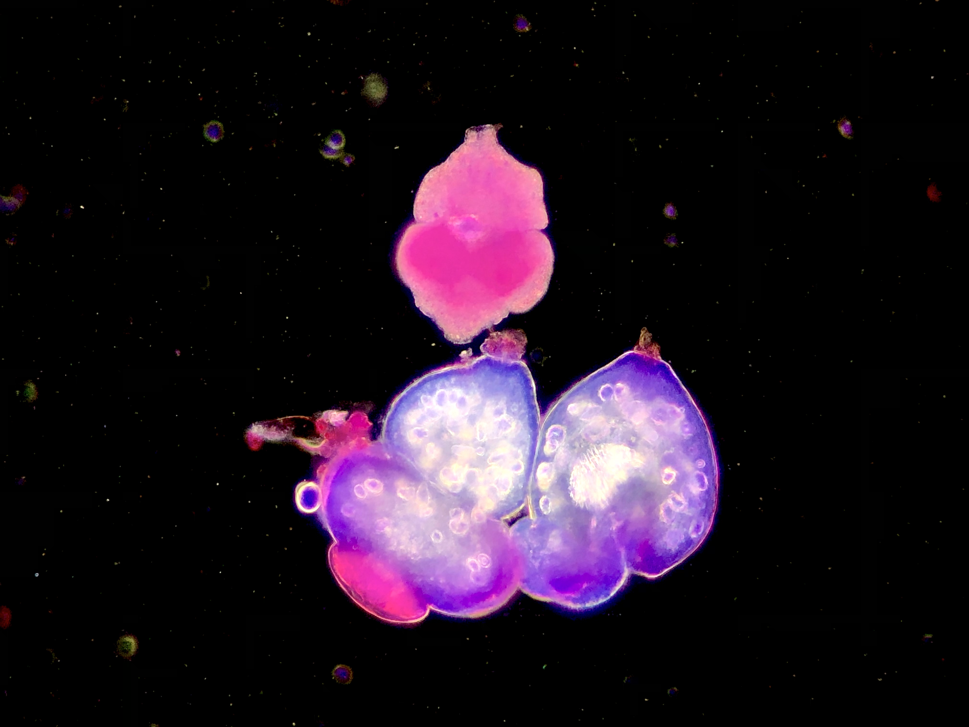 Fine grained Echinococcus protocephalus, stained with SM and photographed in dark field. |
Jurors
 | Geoff Williams, Manager of the Leduc Bioimaging Facility at Brown UniversityGeoff Williams is in his 14th year as manager of the Leduc Bioimaging Facility at Brown University. The opportunity to combine visual arts, science, technology, and mastery of a skill clicked with his discovery of microscopy (electron and light) as an undergraduate at Connecticut College. Geoff transitioned from a graduate program at Michigan State University to running the imaging facility at Central Michigan University before arriving at Brown. Over the past 20-plus years, he has been honing his craft as both an electron and light microscopist, paying more attention to the aesthetics of each image collected than is typically required of a purely scientific investigation. Geoff’s work, under the name Nanoscape, provides a tactile and striking view of samples we may or may not encounter in our day-to-day lives. |
 | Harini Sreenivasappa, Manager of the Cell Imaging Center at Drexel UniversityHarini Sreenivasappa is the manager of Drexel University’s light microscopy core facility, the Cell Imaging Center. She was introduced to microscopy during graduate school at Texas A&M University (TAMU), where she studied the role of microenvironment stimuli on cellular sensing and adapting as it takes place in blood vessel wall remodeling in cardiovascular disease. This led to a PhD in biomedical engineering. She has more than 10 years’ experience working with various microscopy techniques, such as atomic force microscopy (AFM), spinning disk confocal, and total internal reflection fluorescence (TIRF) microscopy. With ASCB’s COMPASS Outreach grant, she created and curated a Traveling Micrographs exhibit showcasing micrographs by TAMU researchers that was free and open to the public. The goal of the series of exhibits was to share research at TAMU with the local community and stimulate interest in imaging science. |
 | Rachid Rezgui, Research Instrumentation Scientist, Microscopy, New York University Abu DhabiRachid Rezgui is a microscopist and an active research scientist. Rachid studied physics at the Leibniz University of Hanover in Germany, then completed his PhD in biophysics at the Ecole Polytechnique in France studying DNA-protein interactions at the single molecule level. He joined the microscopy core facility at New York University Abu Dhabi in 2014, and has since worked with all types of microscopes, including two-photon, super-resolution, confocal, fluorescence lifetime, and widefield. He is involved in all aspects of optical imaging, such as sample preparation, training, acquisition, and post-processing, as well as core facility management. |
 | Urs Ziegler, Managing Director, Center for Microscopy and Image Analysis, ZurichUrs Ziegler studied Biochemistry at the University of Zurich and obtained his Ph.D. from the Institute of Biochemistry in 1996. In 2007, he became Head of the Center for Microscopy and Image Analysis. His research has included the development and introduction of various cryo-electron microscopic methods for the structural study of multicellular organisms, super-resolution light microscopy and new correlative light and electron microscopic methods. In addition to research, he teaches various lectures and hands-on courses in advanced microscopy and image analysis. Urs has also been a member of the Board of the Swiss Society for Optics and Microscopy since 1998. |
 | Yujie Sun, Tenured Professor and Boya Distinguished Professor of Peking UniversityDr. Yujie Sun obtained his bachelor’s and master’s degrees in chemistry from the University of Science and Technology of China and his PhD in chemistry from the University of Pittsburgh. He then joined the University of Pennsylvania School of Medicine as a postdoctoral fellow and worked with an interdisciplinary team to solve the puzzle of how molecular motors work using single molecule fluorescence and manipulation techniques. Dr. Sun is the associate dean of the College of Future Technology (CFT), deputy director of the Nano/Bio Interface Center (NBIC), and deputy chief engineer of the National Multimode Trans-Scale Biomedical Imaging Center. He serves on many professional scientific organizations, including the American Society for Cell Biology (ASCB), the Biophysical Society (BPS), and the Biophysical Society of China (BSC). Dr. Sun has been developing advanced single-molecule imaging and manipulation techniques to study cellular structures and processes. His work is published in five books and more than 100 peer-reviewed papers, and he has undertaken 10 national scientific research projects. |
 | Sarah Ellis, Associate Professor, Centre for Imaging the Tumour Environment (CITE) at the Olivia Newton-John Cancer Research InstituteWith more than 30 years’ experience in all aspects of microscopy, Sarah Ellis has extensive skills in sample preparation for optical and electron microscopy, image data analysis, and the operation and maintenance of widefield, confocal, multiphoton, and biological electron microscopes. Sarah also has broad experience in core facility design and management. She enjoys training researchers and has strong collaborative networks, as evidenced by her inclusion as an author in over 60 publications. Sarah contributes to the scientific community through multiple voluntary roles and is the secretary of the Australian Microscopy and Microanalysis Society and the Victorian representative for Light Microscopy Australia. |
Download Wallpapers
Download the Image of the Year Award 2022 wallpaper package now for free and beautify your screen.
Download the wallpaper package for desktop (ZIP, 136.2 MB)
Download the wallpaper package for mobile (ZIP, 131.4 MB)
Virtual BackgroundDownload a background you can use during virtual meetings. | 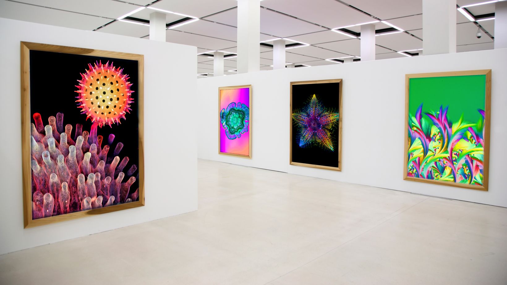 |
Past Winners
|
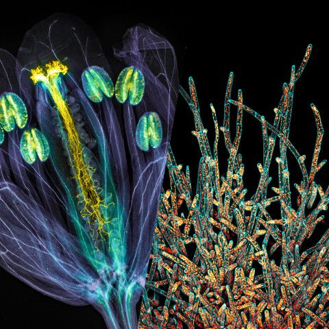
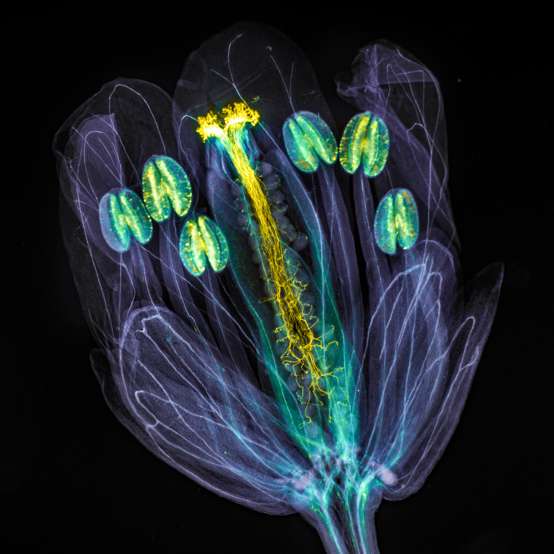
The global winning image was taken by Jan Martinek (Czech Republic). Arabidopsis thaliana flower with pollen tubes growing through the pistil. The flower tissues were chemically cleared to become transparent, while the pollen tubes were stained with aniline blue (yellow fluorescence) in order to be seen. |
Sorry, this page is not
available in your country.
Sorry, this page is not
available in your country.