
Efficient ExperimentsCover a magnification range from 40X to 100X without changing objectives using a 40X X Line objective with a 1.4 numerical aperture and the FV3000 microscope's optical zoom. |  |
  | No Compromise in Confocal Image QualityThe 60X X Line oil immersion objective's improved numerical aperture, image flatness, and chromatic aberration correction deliver outstanding images and reliable data in a wide wavelength range (400–1000 nm). |
|---|
Superior Image Quality at 405 nm ExcitationX Line objectives provide uniform, high-quality images at the 405 nm excitation wavelength over a full field of view (FN18) for precise quantitative analysis, such as counting cell nuclei stained by DAPI. | 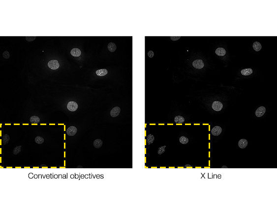 |
 Brain section of Fucci2 transgenic mouse Stitched image of 12X12 images acquired by FV3000 using 60X oil immersion objective (NA1.42) Cyan: DAPI (405nm) Magenta: mCherry ‘561nm) Image Courtesy of: Laboratory for Cell Function Dynamics, RIKEN Center for Brain Science Takako Kogure, Atsushi Miyawaki | Multicolor Panoramic ImageImproved image flatness enables you to acquire superior stitched images with less zooming and in a wide wavelength range starting at 400 nm. |
|---|
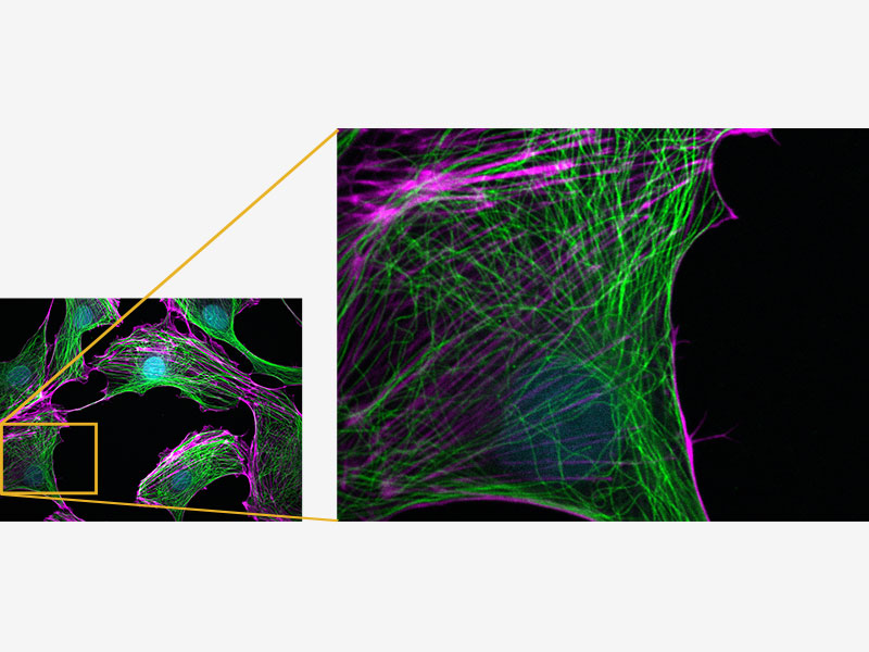
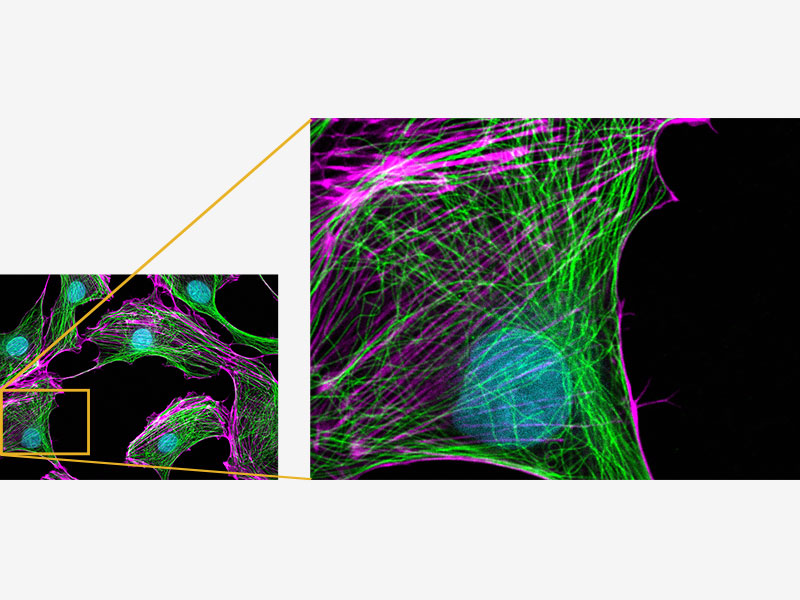
Exceptional Color AccuracyWith chromatic aberration correction from 400 to 1000 nm, X Line objectives deliver accurate multicolor images and quantitative results during colocalization analysis. Image #1: Comparison of chromatic aberration measured by FLUOVIEW FV3000 using TetraSpeck TM Microsphere.Cyan: 405nm excitation, Magenta: 640nm excitation Image #2: HeLa cell labeled by FISH technique CEP17(Spectrum Green), CEP18(Spectrum Orange), Nuclear (DAPI)*1 When observing with conventional objectives, signals located at the bottom of cell nuclear appears outside the nuclear. Scal bar: 2 μm | 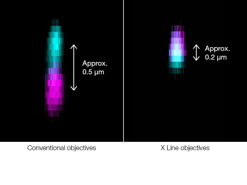 |
*1 Although it became one of the most important cell lines in medical research, it’s imperative that we recognize Henrietta Lacks’ contribution to science happened without her consent. This injustice, while leading to key discoveries in immunology, infectious disease, and cancer, also raised important conversations about privacy, ethics, and consent in medicine.
To learn more about the life of Henrietta Lacks and her contribution to modern medicine, click here.
http://henriettalacksfoundation.org/
Related Products
FV3000
|
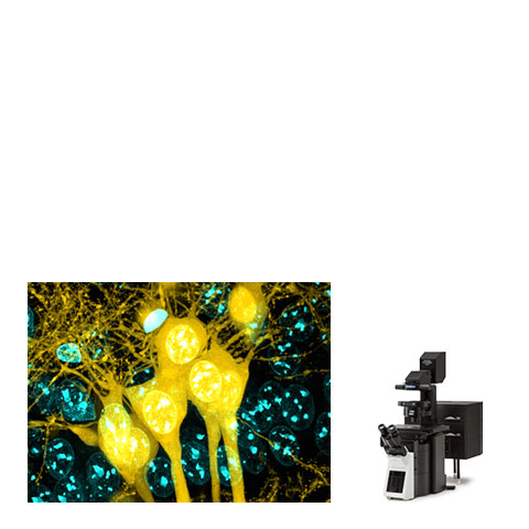
*Banner Image: Fixed brain coronal slice (thickness 1mm) of Thy1-YFP-H mouse Staining: DAPI by ChemScale Mounting solution: SCALEVIEW-SMt (ND=1.49)
MIP image acquired by FV3000 using UPLXAPO60XO(NA1.42) Cyan: DAPI(405nm), Yellow: YFP(488nm)
By courtesy of: Laboratory for Cell Function Dynamics RIKEN Center for Brain Science Tetsushi Hoshida, Hiroshi Hama, Atsushi Miyawaki
Sorry, this page is not
available in your country.
Sorry, this page is not
available in your country.