Three primary functions are performed by actin within the structure of the cell. Actin allows cell motility, the ability to move spontaneously. Secondly, it forms the most dynamic of the three subclasses of the cytoskeleton, which gives mechanical support to cells, and hardwires the cytoplasm with the surroundings to support signal transduction. Thirdly, in muscle cells as well as non-muscle cells, actin helps generate force to support muscle contraction, vesicle movement, and other transport processes. In the digital video presented above, embryonic rat thoracic aorta fibroblast cells (A7r5 line) are expressing a fusion of JRed fluorescent protein to human beta-actin.
Video 1 - Run Time: 60 Seconds
Video 2 - Run Time: 51 Seconds
Video 3 - Run Time: 57 Seconds
Video 4 - Run Time: 50 Seconds
There are two types of actin bundles: parallel and contractile. In parallel bundles, the actin-bundling protein fimbrin spaces the filaments about 14 nanometers apart. Parallel bundles are responsible for supporting a cell’s microvilli, the structures that increase the cell’s surface area. In vertebrates, the actin-bundling protein villin is almost entirely found in the microvilli of intestinal cells. In contractile bundles, the actin-bundling protein actinin separates each filament by about 40 nanometers. This increase in distance allows the motor protein myosin to interact with the filament, enabling deformation or contraction. In the digital video presented above, embryonic rat thoracic aorta fibroblast cells (A7r5 line) are expressing a fusion of JRed fluorescent protein to human beta-actin.
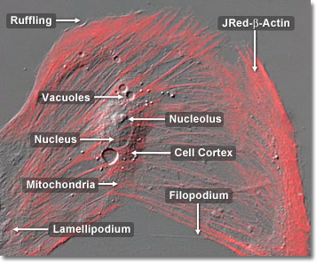
Although it can be found throughout the cell, the highest concentration of actin is in the region close to the plasma membrane. In overall terms, actin can be likened to a peripheral layer around the cell, like an elastic stocking or a muscular skin. This layer is known as the cell cortex and gives the outer surface of the cell mechanical strength and the ability to move. Because of its actin cortex, an animal cell can produce extensions of its surface, crawl, engulf particles, and deform its external shape. All of most distinctive features of higher animals, such as their complex internal anatomy, their immunity to infection and their complex nervous systems, depend on the actin cortex. In the digital video presented above, embryonic rat thoracic aorta fibroblast cells (A7r5 line) are expressing a fusion of JRed fluorescent protein to human beta-actin.


