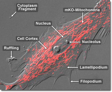
The mitochondrion is different from most other organelles because it has its own circular DNA (similar in structure to the DNA of prokaryotes) and reproduces independently of the cell in which it is found; an apparent case of endosymbiosis. Scientists hypothesize that millions of years ago small, free-living prokaryotes were engulfed, but not consumed, by larger prokaryotes, perhaps because they were able to resist the digestive enzymes of the host organism. The two organisms developed a symbiotic relationship over time, the larger organism providing the smaller with ample nutrients and the smaller organism providing ATP molecules to the larger one. Eventually, according to this view, the larger organism developed into the eukaryotic cell and the smaller organism into the mitochondrion. In the digital video presented above, normal Gray fox lung fibroblast cells (FoLu line) expressing a fusion product of monomeric Kusabira Orange fluorescent protein fused to a mitochondrial targeting sequence are imaged with a combination of confocal fluorescence and differential interference contrast (DIC) illumination. Note the paths of individual mitochondria as the cell(s) slowly migrate across the glass coverslip.
Video 1 - Run Time: 47 Seconds
Video 2 - Run Time: 51 Seconds
Video 3 - Run Time: 58 Seconds
Video 4 - Run Time: 67 Seconds
Video 5 - Run Time: 47 Seconds
Video 6 - Run Time: 51 Seconds
Video 7 - Run Time: 30 Seconds
Video 8 - Run Time: 59 Seconds
Video 9 - Run Time: 27 Seconds
Video 10 - Run Time: 57 Seconds
Video 11 - Run Time: 66 Seconds
Video 12 - Run Time: 36 Seconds
Video 13 - Run Time: 49 Seconds
Video 14 - Run Time: 53 Seconds
Video 15 - Run Time: 65 Seconds
Video 16 - Run Time: 52 Seconds
Video 17 - Run Time: 47 Seconds
Video 18 - Run Time: 05 Seconds
Video 19 - Run Time: 61 Seconds
Video 20 - Run Time: 48 Seconds
The elaborate structure of a mitochondrion is very important to the function of the organelle. Two specialized membranes encircle each mitochondrion, dividing the organelle into a narrow intermembrane space and a much larger internal matrix, each of which contains highly specialized proteins. The outer membrane of a mitochondrion contains many channels formed by the protein porin and acts like a sieve, filtering out molecules that are not needed. Similarly, the inner membrane, which is highly convoluted so that a large number of infoldings called cristae are formed, also allows only certain molecules to pass through it and is much more selective than the outer membrane. To make certain that only those materials essential to the matrix are allowed into it, the inner membrane utilizes a group of transport proteins that will only transport the correct molecules. Together, the various compartments of a mitochondrion are able to work in harmony to generate ATP in a complex multi-step process. In the digital video presented above, normal Gray fox lung fibroblast cells (FoLu line) expressing a fusion product of monomeric Kusabira Orange fluorescent protein fused to a mitochondrial targeting sequence are imaged with a combination of confocal fluorescence and differential interference contrast (DIC) illumination. Note the paths of individual mitochondria as the cell(s) slowly migrate across the glass coverslip
Mitochondria convert oxygen and other nutrients into adenosine triphosphate (ATP), the chemical "currency" of the cell. These rod-shaped organelles are considered the power generators of the cell, driving metabolic activity. The conversion process responsible for the formation of ATP inside mitochondria is an aerobic activity requiring oxygen to proceed. Higher animals would not be able to obtain enough energy from anaerobic respiration (without oxygen) because it is much less efficient. All complex animals need large amounts of energy to exist, and the aerobic respiratory activity of mitochondria promotes the production of 15 times more ATP which can then be easily converted into usable energy. In the digital video presented above, normal Gray fox lung fibroblast cells (FoLu line) expressing a fusion product of monomeric Kusabira Orange fluorescent protein fused to a mitochondrial targeting sequence are imaged with a combination of confocal fluorescence and differential interference contrast (DIC) illumination. Note the paths of individual mitochondria as the cell(s) slowly migrate across the glass coverslip.


