As a protein involved in crosslinking actin filaments, alpha-actinin may be one of the proteins involved in attachment of the actin cytoskeletal network to the plasma membrane in a wide spectrum of cell types.
Video 1 - Run Time: 59 Seconds
Video 2 - Run Time: 65 Seconds
Video 3 - Run Time: 55 Seconds
Video 4 - Run Time: 58 Seconds
Video 5 - Run Time: 60 Seconds
Video 6 - Run Time: 12 Seconds
Video 7 - Run Time: 18 Seconds
Video 8 - Run Time: 20 Seconds
Video 9 - Run Time: 19 Seconds
Video 10 - Run Time: 59 Seconds
Video 11 - Run Time: 60 Seconds
Video 12 - Run Time: 51 Seconds
Video 13 - Run Time: 60 Seconds
Video 14 - Run Time: 36 Seconds
Video 15 - Run Time: 56 Seconds
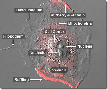
A member of the spectrin superfamly, alpha-actinin is an actin crosslinking protein that forms antiparallel homodimers in a rod-like structure with one actin-binding domain on each side of the rod. In addition to its binding to actin, alpha-actinin also associates with a number of other cytoskeletal proteins including titin, zyxin, vinculin, and alpha-catenin, as well as cytoplasmic signaling proteins, such as phosphatidylinositol 3-kinase, Rho effector kinase, and the beta-integrins. In the digital video presented above, a normal rabbit kidney epithelial cell (RK-13 line) expressing a fusion of mCherry fluorescent protein with human alpha-actinin is observed traversing the coverslip in a live-cell imaging chamber using fluorescence and differential interference contrast (DIC) illumination. Note the high levels of labeled alpha-actinin present in the lamellipodia, which continually undergo wave-like motions.
In addition to its role in linking the actin cytoskeletal network to focal adhesions, alpha-actinin is also thought to be involved in signaling pathways. This ubiquitous protein also has a number of other molecular partners (in addition to actin), and is present in multiple subcellular regions, including cell-cell contact sites, protrusions, lamellipodia, and stress fibers. In the digital video presented above, a normal rabbit kidney epithelial cell (RK-13 line) expressing a fusion of mCherry fluorescent protein with human alpha-actinin is observed traversing the coverslip in a live-cell imaging chamber using fluorescence and differential interference contrast (DIC) illumination. Note the high levels of labeled alpha-actinin present in the lamellipodia, which continually undergo wave-like motions.


