Oblique Illumination Refractive Index Determination
Oblique Illumination Refractive Index Determination - Java Tutorial
Oblique illumination is sometimes utilized as an alternative to the Becke line test to determine whether the refractive index of a specimen is higher or lower than that of the surrounding medium. This interactive tutorial explores how variations in the refractive index of a specimen and its surrounding medium alter visibility in the microscope when utilizing oblique illumination techniques.
The tutorial initializes with a specimen having a refractive index of 1.15 positioned in a surrounding medium of refractive index 1.33 (representing water or lightly buffered aqueous saline solution), and being illuminated with off-axis light rays originating from the lower left-hand side of the tutorial window. In order to operate the tutorial, translate the Specimen (refractive index value) slider between values of 1.0 and 1.5. As the slider is moved from right to left, the specimen appearance is altered in the Eyepiece View window. To change the refractive index of the surrounding medium, move the Surround (refractive index value) slider to the right or left (this slider also has a range of refractive indices between 1.0 and 1.5). Translating the Surround slider will also affect specimen appearance and visibility, as discussed below. The two sliders are interactive and positioning of one slider will affect the range of motion of the other.
In situations where the specimen is mounted in a medium of lower refractive index, shading that results from the anaxial illumination will appear on the side opposite to that from which the light enters the specimen, and vice versa, as illustrated in Figure 1. For both diagrams presented in Figure 1, two equal sized oblique light rays are depicted entering the specimen through the surrounding medium at the same angle of incidence. At point A on the left-hand diagram, the light is spread over a larger area of the specimen than at point B, so that the area near point A on the specimen appears darker than the area near point B. Under these conditions, one side of the specimen will appear shaded or somewhat darker than the other side when viewed through the microscope eyepieces (represented by points A' and B' in the upper left portion of Figure 1). This is the case when the specimen refractive index is higher than that of the surrounding medium.
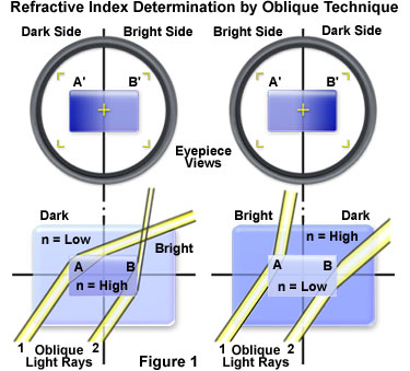
The opposite effect occurs when the specimen has a lower refractive index than that of the surrounding medium (see the right-hand side of Figure 1). In this case, the shaded or darker side of the specimen will be on the side that is nearest to the oblique light sector stop. When the specimen and the surrounding medium have identical refractive indices, then the specimen will be transparent (or invisible) and will have no refractive effects on the oblique illumination.
The sensitivity of this refractive index determination technique is highly dependent on the condenser focal length, the iris diaphragm position, and the geometry of sector stops (if employed). In general, the best results are obtained when the condenser is carefully focused and an even field of illumination is achieved.
Sorry, this page is not
available in your country.


