Total Support for Assisted Reproductive Technology (ART) Research
Olympus microscopy solutions for assisted reproductive technology (ART) research applications range from stereoscopic microscopes used for oocyte cleaning, oocyte freezing, and embryo thawing observation to upright microscopes for sperm observation, to inverted microscopes that support ICSI/IMSI workflow and additionally embryo manipulation using lasers.
|
|
|
|

Sperm Observation
System Microscope BX53 System Microscope BX53* |
Biological Microscope CX43 Biological Microscope CX43* |
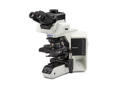

Sperm is typically observed by contrasting bright and dark on the sample, using phase contrast observation under an upright microscope. A common limitation of these microscopes is that their light source is not bright enough to properly verify sperm mobility. Olympus’ BX53 microscope system has a high-luminosity LED that enables users to clearly observe the mobility of sperm. In addition, since the LED light source generates low heat, damage to the sample is diminished. The ergonomic observation tubes and negative phase contrast objective for longer and clearer sperm observations help ensure comfortable microscope operation. The CX43 biological microscope, which is an entry-level model, is designed with low stage so users can keep their arms on the desk, helping reduce fatigue during long observation sessions. It is compact model with a small footprint to save space in the lab.
Comment by Kato Ladies Clinic
We use the upright BX53 microscope mainly for sperm counting and morphological observation. Sperm are counted by phase contrast observation using the SMAS (sperm motility analysis system). Olympus upright microscopes have a sufficient amount of light that does not interfere with measurement. For sperm morphology evaluation, unstained specimens are prepared by our own method and observed using 600X dry objectives. The outline of the sperm can be clearly seen, and the contrast adjustment enables us to observe and evaluate the shading of the sperm head at the same image-quality level as stained specimens. Furthermore, incorporating differential interference contrast (DIC) observation increases the clarity and improves the accuracy of sperm morphology evaluation.
Research content of this application note was prepared with the collaboration of the following user:
Dr. Kazuo Uchiyama
Kato Ladies Clinic
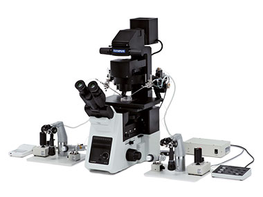
ICSI/IMSI
 |
| ||
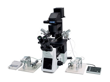
Inverted Microscope IX3-ICSI/IMSI | |||
When performing intracytoplasmic sperm injection (ICSI) in an oocyte, polarization observation should be used to observe the spindle to confirm that it is in the metaphase II stage. This indicates it has become a mature oocyte, and also helps to make sure that it is in the right position to avoid damage during injection. Olympus’ IX3 inverted microscope series with its polarization technology enables users to clearly observe the appearance of the spindle. Also, Olympus' relief-contrast allows the observation of 3-dimentional oocytes in plastic dishes, which enables users to check the condition of oocyte zona pellucida.
During ICSI and IMSI, multiple observation methods and lens magnifications are used, making microscope operation more complex and less efficient and increasing the level of stress and fatigue for the user. The Olympus IX3 semi-motorized system has an ICSI-dedicated hand switch enabling the user to instantly change the observation method and magnification to suit each step in process with the touch of a button, saving time and reducing fatigue. This functionality can even eliminate certain steps of the traditional process. It also means that users don't have to take their eyes off the eyepieces, and consequently can focus on delicate manipulator operations.
With IMSI, which is a method of injecting sperm selected by morphological characteristics into oocytes, the shape, size, and number of vacuoles on the sperm head are confirmed at high magnification. This is supported by the IX3 series’ differential interference contrast (DIC) observation.
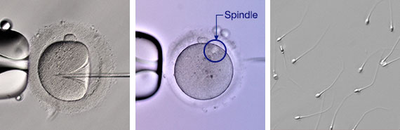
| ICSI (Relief contrast) | Spindle (Polarizing observation) | Sperm (DIC) |
Comment by Kato Ladies Clinic
Sperm used for ICSI are selected by IMSI using high-magnification differential interference contrast observation. Since implementing this method, the probability of selecting normal morphological sperm has increased compared with conventional observation using 200X or 400X objectives. Correctly choosing sperm with the normal morphology reduces the DNA fragmentation rate which may be beneficial for patient safety, for example, by lowering the miscarriage rate.
Visualization of the spindle by polarizing observation is effective for correctly measuring the timing to perform ICSI. Even when the oocyte has extruded the first polar body, successful fertilization is less likely if ICSI is performed in an oocyte without meiotic spindle visualization. Since we are able to visualize the meiotic spindle at the time of ICSI, our fertilization rate has significantly improved.
The Olympus IX83 inverted microscope can seamlessly perform a series of observations with differential interference contrast (DIC), polarization, and relief contrast, and the IX73 semi-motorized model can switch between polarization and relief contrast by the remote controller. The Olympus IX83 and IX73 are easy-to-use devices for users.
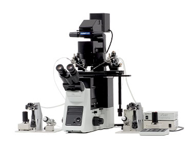
Laser Manipulation of Embryos
Inverted Microscope IX3-ICSI/IMSI |
As a post-fertilization process, detailed embryo examination has an increasing number of applications, such as assisted hatching and preimplantation genetic testing for aneuploidy (PGT-A). Olympus’ IX3 inverted microscope series provides a high-quality, clear view of the oocyte by combining* the microscope with a laser system and providing a dedicated remote switch to easily optimize the contrast.
*The combination and configuration with the microscope will differ depending on the laser manufacturer.
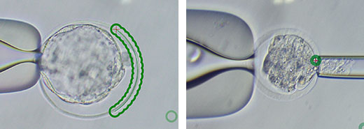
Laser manipulation in assisted hatching and PGT-A |
*Samples and images data are courtesy of Cooper Surgical Inc.
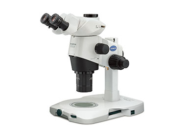
Fertilization Check of Oocytes and Embryos
Research Stereo Microscope System SZX16 Research Stereo Microscope System SZX16* |
Fertilized Embryo |
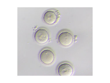
Oocyte cleaning, fertilization checks, embryo grade confirmation, and observation during oocyte freezing and thawing are performed under a stereo microscope. Since it is difficult to see the oocyte and embryo, it takes time to find and judge the maturity level, placing a burden on the user. To help ensure the user’s comfort, Olympus SZX stereo microscope series has adjustable eye points and uses an observation tubes with a convergence angle, which alleviates eye fatigue. There are also several ergonomic accessories available for the series, such as an observation tube that can incorporate a clean bench because the mounting position of the main body and eyepiece is longer than usual.
In addition, the SZX2-ILLTQ/S transmitted light illumination base can provide appropriate contrast and uniform illumination from low to high magnification, so the condition of oocytes and embryos can be checked quickly and work efficiency can be improved. Olympus’ stereo microscope SZX series provides a comfortable microscope workspace and contributes to reducing user's fatigue and improving work efficiency. SZX2-ILLTQ/S also enables users to choose the appropriate observation method and contrast from 9 types of cartridges, depending on the application and user's preference.
Comment by Kato Ladies Clinic
Stereo microscopes are used in a wide range of applications such as oocyte denuding, oocyte and embryo observation, and vitrification-warming. Brightfield, oblique, and darkfield observation methods are used according to the laboratory procedures. Olympus' stereo microscope enables you to quickly switch all observation areas from 0.7X to 11.5 X magnification in focus, which improves work efficiency. Since optimal contrast can be selected from three fine-adjustable contrast levels, the observation of oocytes and embryos has become much easier.
We use a 0.8X objective lens and combine an ergonomic trinocular barrel (SZX-LTTR) with an intermediate magnification of 1.25x to ensure a large working space under the objective lens. As a result, work efficiency for oocyte and embryo observation and manipulation has improved. In addition, since the light source uses LEDs, it is less likely to deteriorate and is maintenance-free, which is also highly valued.
* For research use only.
Produtos usados nesta aplicação
Sorry, this page is not
available in your country.