The skin is the first line of defense and the immune system’s biggest barrier for combating pathogens. Being able to accurately characterize and identify immune cell subtypes, tissues structures, and cell distribution in the skin under steady-state conditions provides a powerful tool for understanding the first immunological strategies and biological processes that occur in the presence of pathogens. In this webinar we will review technical aspects involved in the experimental process and explore how complementary imaging technologies might assist us to better understand the immune system.
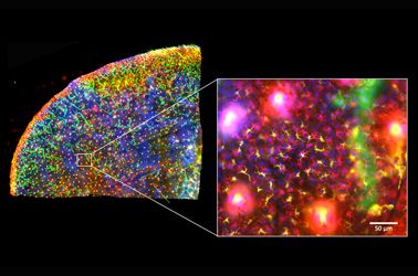
The presentation is divided into three parts. First, an introduction of the Hugh Green Cytometry Centre will be presented and an overview of the histology and bioimaging technological platforms available. Second, the multiplexing methodology will be discussed, where several topics need to be considered for the design and development of a successful polychromatic panel for microscopy. Finally, preliminary results from a research project will be presented that constitutes part of a diploma program from The Royal Microscopical Society. The project focuses on the identification of immune cell types in the whole mount skin in relation to tissues structures (e.g., blood vessels and lymphatic network). It also centers on the immune cells’ distribution in the tissue as a first barrier of defense against pathogens.
Presenter: Alfonso J. Schmidt
Senior Staff Scientist, Malaghan Institute of Medical Research
Alfonso has a decade of experience working in a Shared Resource Lab (SRL) with a vast knowledge in histology, fluorescent microscopy, and image analysis. His work has been focused in maximizing the capabilities of the equipment available and in creating technical protocols and training modules for the scientific community. Currently, Alfonso oversees the Histology and Bioimaging Facility as part of the Hugh Green Cytometry Centre (HGCC) at the Malaghan Institute of Medical Research. Wellington, New Zealand.