
October has come and gone, and with it the spooky energy of ghosts, witches, and zombies. This month, our Instagram followers shared a great deal in common with zombies, as both clearly love brains!
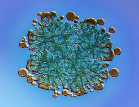
“Algal bloom is a common problem during summers in Stockholm. A couple of weeks ago, I collected these green algae during a bloom close to where I live. It is a microscopic colony of a green algae called "Botryococcus Braunii.” It measures about 140-micrometers across. Colonies are held together by a lipid (oily) biofilm, and you can see old droplets around the edge of the colony. Typically, around 30–40% of the dry weight of a colony is oil. Blooms of Botryococcus braunii have been shown to be toxic to other micro-organisms and fish but they may have other applications than being a source of food. Botryococcus braunii has great potential for algaculture because of the hydrocarbons it produces, which can be chemically converted into fuels. Botryococcus oils can be used as feedstock for hydrocracking in an oil refinery to produce octane (gasoline), kerosene, and diesel. Photographed in DIC.”
Image captured with an Olympus UplanXAPO 60X objective using oil immersion. Caption and image courtesy of Håkan Kvarnström.
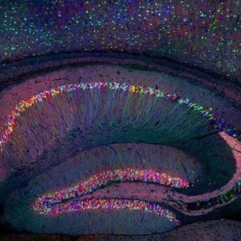
The last week of October, we celebrated Neuroscience Week 2020 with a series of tech talks, product demonstrations, and research presentations all focused on the brain. The above image is a great example of the Brainbow technique, one of our favorites and a colorful way to capture a brain image, with each neuron labeled to express a distinct color.
Image captured by Tamily Weissman.
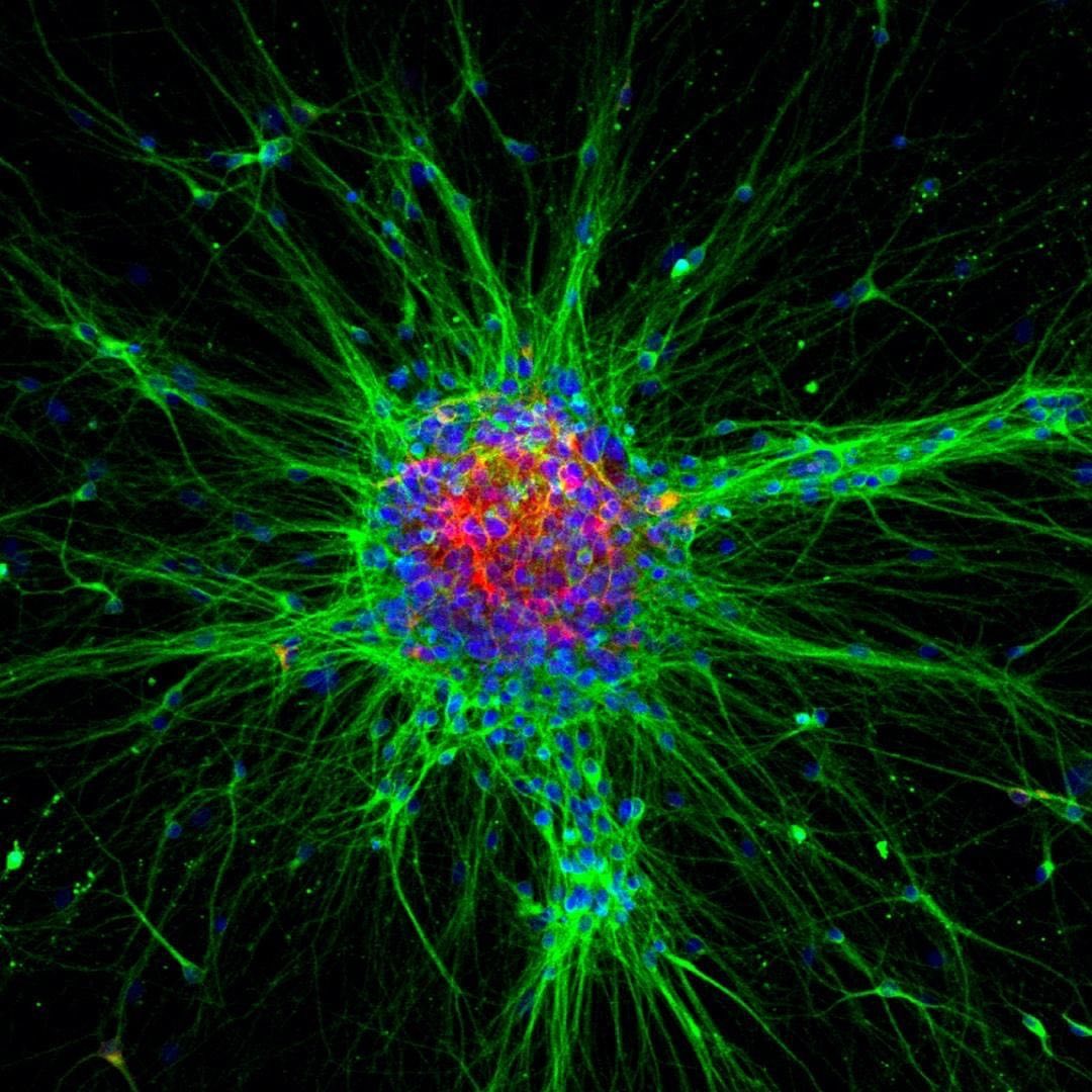
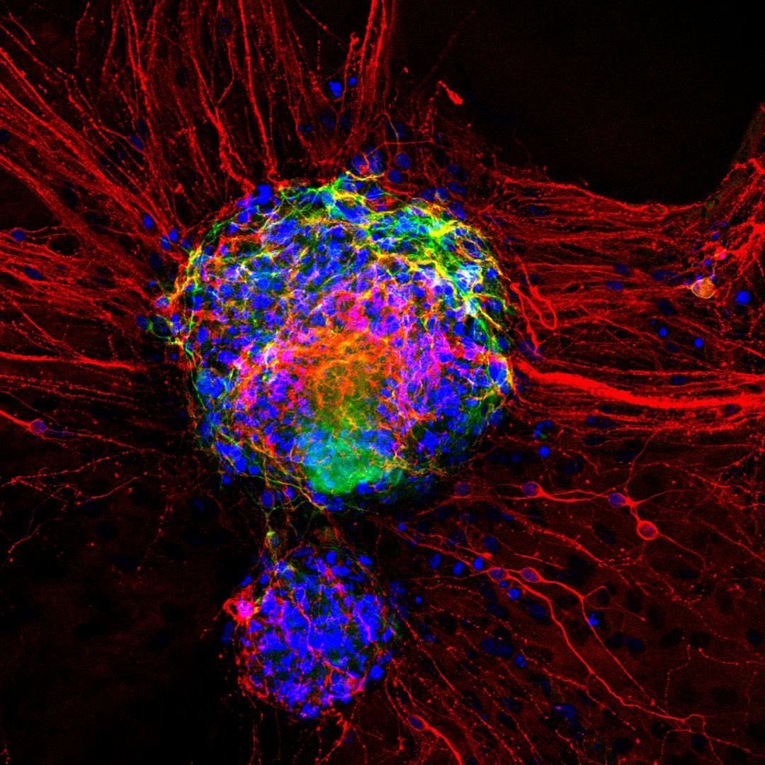
During Neuroscience Week, we featured a mini Instagram Takeover from researchers at our Discovery Center at the Douglas Mental Health University Institute. This “TBT” to their 2019 Olympus Discovery Center Imaging Contest takes another look at the first, second, and third place (L to R) images from research students at McGill University.
Image 1: First Place—This image visualizes cortical neurons differentiated from urine-derived induced pluripotent stem cells. Neurons are labeled for lineage-specific markers MAP2 (green) and TUJ1 (red), in addition to nuclei staining with DAPI (blue).
Image courtesy of Nuwan Hettige. Captured on an Olympus FV1200 laser scanning confocal microscope.
Image 2: Second Place—This image captured a sphere of dopaminergic neurons one day after attaching to a coverslip.
Image courtesy of Scott Bell. Captured on an Olympus FV1200 laser scanning confocal microscope.
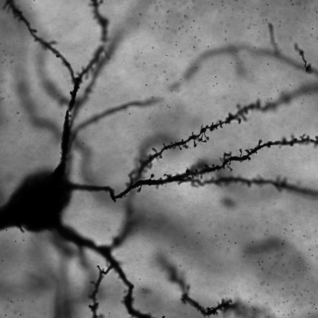
Image 3: Third Place—This image shows Golgi-Cox stained neurons in a brain slice from a female rat medial prefrontal cortex.
Image courtesy of Mary Loka. Captured with brightfield microscopy using an Olympus BX63 widefield microscope.
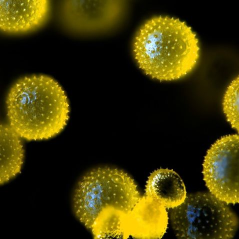
Did you know October 26 was National Pumpkin Day in the US? To celebrate, this image shows an important function of pumpkin growth—pollination! This image shows the magnified pollen grains of a pumpkin flower. Pumpkin flowers are not self-pollinating, so they require assistance from bees or human-induced pollination to improve the quality of the fruit (and, in turn, the quality of our pies for the autumn season)!
Image courtesy of Johann Swanepoel. Captured on an Olympus BX53 microscope at 200x magnification.
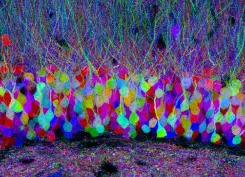
These pastel neurons, captured with confocal microscopy, are located in the hippocampus of a "Brainbow" mouse brain. This is another stunning example of this scientific and artistic technique.
Image courtesy of Dr. Jean Livet.
To see more images like these, be sure to follow us on Instagram at @olympuslifescience!
Interested in sharing your own images?
Visit our image submission site.
Submissions are now also being accepted for our 2020 Global Image of the Year Award Competition. Click here to learn more and see and how you can win an SZX7 stereo microscope with DP27 digital camera or CX23 microscope.
Related Content
Missed last month’s top images? See the best of September
5 Practical Ways to Accelerate Your Microscopy Experiments
Combining Passions for Science and Art—Meet the 2019 IOTY Americas Regional Winner
