Innovative Cell Observation Technology which Enabled CM20's Compact Design
An Innovative Device Makes Cell Culture Smarter
The Olympus CM20 incubation monitoring system (Figure 1) enables you to remotely observe the health of cell cultures in an incubator without entering the clean room. Its thin, compact design fits inside most standard-sized incubators, eliminating the need to prepare a special incubator. It also has a flat-top surface, enabling you to place and observe large, tall vessels such as multi-layer flasks. To achieve this compact design, we developed an innovative, patented optical system known as epi-oblique illumination.
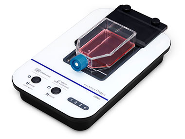
Figure 1. CM20 incubation monitoring system
Designing a Compact Cell Culture Monitor
Incubation monitoring devices that can observe the whole vessel are typically large and occupy considerable space inside the incubator. As a result, a special environment or a new incubator must be prepared.
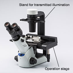
For instance, a conventional method to observe label-free cell cultures involves using an inverted microscope with phase contrast or differential interference contrast. This method requires the observation optical system and illumination optics to be positioned on each side, with the sample in between. Additionally, the operation stage must enable observation of the whole vessel. As a result, the system is large and complicated (Figure 2). It takes up valuable space in the incubator and also makes the cleaning process more time-consuming. Olympus set out to create a compact, thin device that saves space in incubators. To do so, we developed a new observation method: epi-oblique illumination. |
Figure 2. Inverted microscope for cell culture |
The Technology Behind Epi-Oblique Illumination
Our epi-oblique illumination technology made it possible to observe transparent samples and enabled us to miniaturize our incubation monitoring system (Figure 3). This technology is unique because oblique lighting uses reflected light from the vessel’s top (e.g., top of flask, lid of well, or petri dish). As a result, the observation optical system and the illumination optics, including the light source, can be placed inside the device. This setup eliminates the need for a conventional transillumination device with a stand (Figure 3) and enabled us to make the device’s top surface flat. The CM20 device is the industry’s thinnest* incubation monitoring system at only 55 mm (2.16 in.) (without a vessel holder). Its compact size enables users to install it in their current incubator—even with limited space (Figure 4).
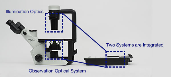
Figure 3. Comparing the size of the conventional cell culture microscope (left) and the CM20 system (right). From this side view, you can clearly see how compact the CM20 system is compared to a conventional setup
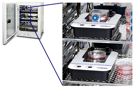
Figure 4. The CM20 system installed inside the incubator
Device Configuration and Captured Images
Transmitted illumination is generally used for oblique lighting, but our epi-oblique illumination casts light from the objective lens side. Lights irradiated from the light source on the objective lens side reflect on the vessel top. Reflected light enters the sample at the angle above the optical axis of objective lens, and the transmitted light that travels through the sample is imaged with the objective lens (Figure 5). Our CM20 system always aligns the incident angle to the sample with the objective’s NA. This creates the same conditions as oblique lighting, enabling you to easily view transparent samples like cells with contrast (Figure 6).
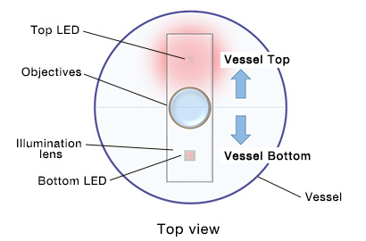 | 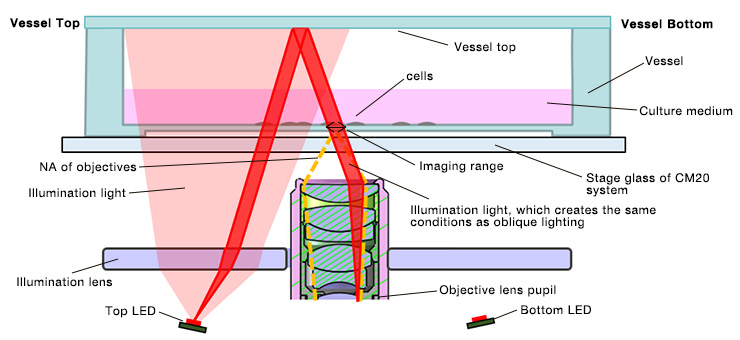 |
Figure 5. Configuration of epi-oblique illumination (side view)
iPS cells (feeder-free) |
MSC |
MEF cells |
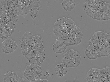
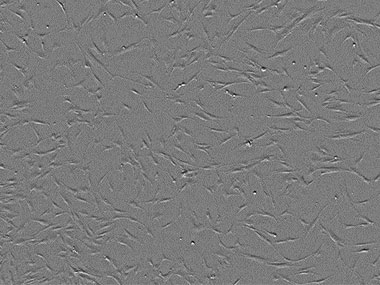
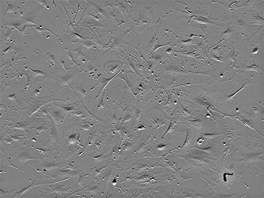
Figure 6. Images captured using epi-oblique illumination
Compatible with Various Types of Vessels
Our new illumination optics are compatible with various vessel types. The illumination light is near the outside for tall vessels and near the inside for short vessels (Figure 7). To prevent the vessel wall surface from affecting the lighting conditions, light sources are arranged in two directions: top and bottom. Stable lighting conditions are maintained by illuminating from the bottom light source when imaging the vessel’s top half and by illuminating from the top light source when imaging the vessel’s bottom half (Figure 8).
Another advantage of epi-oblique illumination is that observation is possible when several vessels are stacked because it uses the reflected light from the vessel top. Additionally, observation with large vessels, such as multi-layer flasks, is possible, overcoming a common challenge with conventional microscopes (Figure 9).
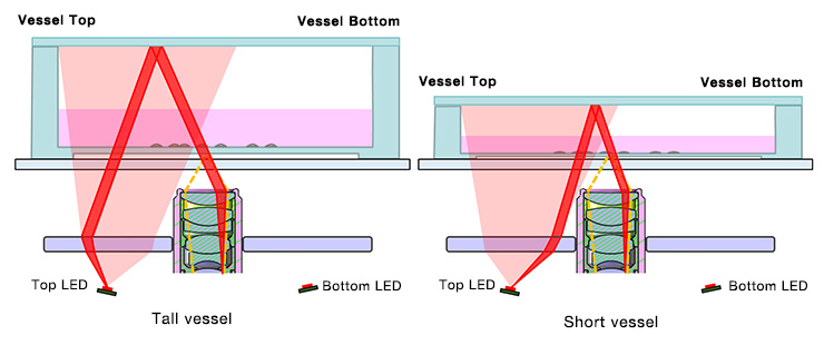
Figure 7. Illumination optics are compatible with tall and short vessels (side view)
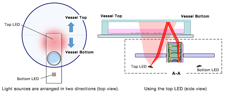
Figure 8. Imaging the bottom half of the vessel
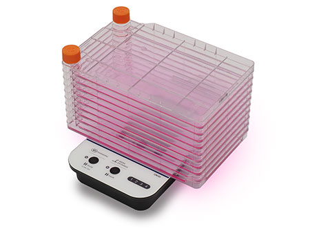
Figure 9. Multiple cell culture vessels stacked on a CM20 monitor
Conclusion
By developing epi-oblique illumination, Olympus has succeeded in engineering a compact, flat-top monitoring device that can observe a vessel’s whole surface. This innovation can help improve cell culture efficiency while saving space in the incubator.
Fine print: *As of April 2020 according to Olympus.
Author
Masaru Mizunaka
Scientific Solutions Product &Technology
Optical System Dev.,CRDM
Olympus Corporation
Sorry, this page is not
available in your country.