Digitizing Slides Using a Manual Microscope and Digital Camera
The Growing Importance of Digitizing Slide Samples
It is becoming more important to increase the efficiency of lab operations by digitizing slide specimens, a practice known as whole slide imaging (WSI). Whole slide imaging has many productivity benefits since it does not require you to set up a microscope to observe a specimen. Instead, you can view virtual slide images from your PC, where you can change the observation position and magnification, as well as observe a sample simultaneously with remote colleagues.
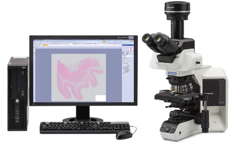
While fully motorized virtual slide scanners can streamline the WSI process, not every researcher has the budget, bench space, or data capacity to own one. As an alternative, you can combine Olympus’ BX53 manual microscope with a DP74 digital camera to perform cost-effective whole slide imaging in a confined workspace.
Digitizing Slides Using Manual Whole Slide Imaging
Manual whole slide imaging (mWSI) creates digital image data of a whole specimen without using a motorized stage. The mWSI approach combines Olympus’ BX53 manual microscope, DP74 digital camera, and cellSen software. It is an effective method for creating stitched images at a low magnification. As you move the stage manually, the acquired images will stitch together in real time. The entire specimen image can be taken within an arbitrary range. For instance, a 15 mm2 specimen area can be scanned in about 30 seconds using a 4X objective lens.
Common Applications for Manual Whole Slide Imaging
Case 1. Group discussions
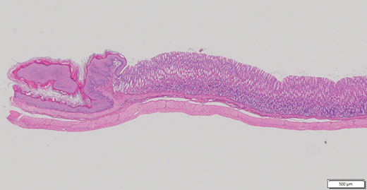
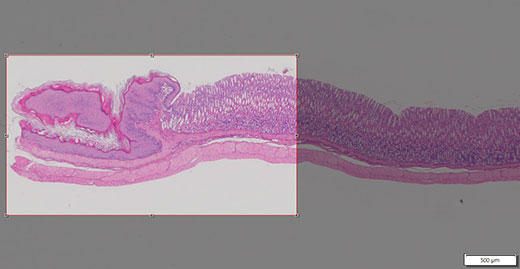
In certain situations, like a study group, you may need to show the entire specimen in both high and low magnification. With manual whole slide imaging, you can capture an image of the entire specimen using a low-magnification objective (Figure 1). The group can discuss by observing the virtual slide image on a screen rather than setting up and using a microscope. mWSI is also useful for discussing sample images taken at high magnification since you can see where the image is located on the overall sample slide (Figure 2).
Related VideosFigure 1: A low-magnification image of a specimen on the sample | Related VideosFigure 2: You can easily find the image location |
Case 2. Images for academic conferences and research papers
Academic conference posters and papers often need to display a wide range of images. Yet, it can be difficult to capture the entire sample, even with a low-magnification objective. Using manual whole slide imaging, you can seamlessly stitch multiple areas together to acquire a clear overview image (Figure 3), then trim the tiled image to capture a certain area (Figure 4).
Fig. 3: Wide overview image of a sample |
Fig. 4: You can trim the overview image to capture the area of interest |
Case 3. Digital archive
In the past, fully motorized virtual slide scanners were necessary if you wanted to capture an entire specimen as a virtual slide image. But with manual whole slide imaging, you can create a digital archive using less data simply by adding a DP74 digital camera and cellSens software to your BX53 microscope.
For instance, some specimens, like cell clusters, require you to capture the multifocal plane. This can be easily done by taking a video while manually shifting the focus position, making it possible to obtain the necessary information in a smaller file size.
Related VideosFigure 5: Image of a cell cluster |
Case4. Remote consultation
Remote observation discussions, which are common in education, require the presenter to electronically share sample images captured under the microscope. However, it can be a difficult and time-consuming task to set up a microscope for a video conference. By digitizing the whole specimen in advance using manual whole slide imaging, you can easily review a sample with remote colleagues and consultants.
Improve Observation with Manual Whole Slide Imaging
Using our manual whole slide imaging method that combines Olympus’ BX53 manual microscope, DP74 digital camera, and cellSens software, you can more efficiently observe specimens at a lower cost than with motorized virtual slide scanners.
Products related to this application
Sorry, this page is not
available in your country.