
Over at the Olympus Life Science Instagram account, we share a wide variety of images captured under the microscope. In microscopy, science and art intersect to create incredibly beautiful colors and structures. Each month, we’ll have a look back blog post and see which images have caught the eye of our followers.
The top five images for October are:
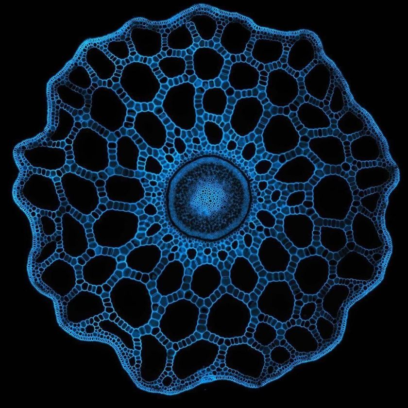
What you're looking at here is one of the few examples of a "living fossil." Equisetum, commonly known as horsetail grass, is the only living genus in a class of organisms whose origins date back over one hundred million years! Equisetaceae is a family of vascular plants that reproduce by spores rather than seeds.
Image courtesy of Olivier Leroux.
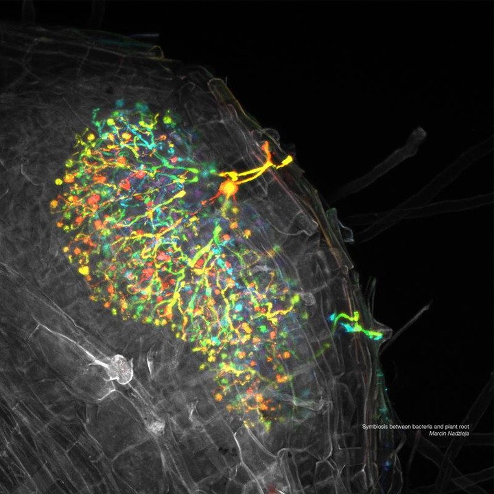
This image from our European 2018 Image of the Year contest captures the symbiosis between bacteria and plant roots. Nitrogen-fixing bacteria called rhizobia enter the roots of leguminous plants via tube-like structures called infection threads. Initially, few infection threads branch to form a complex network, enabling for colonization of larger areas of the root, as depicted in the image.
Image courtesy of Marcin Nadzieja, 2018 Image of the Year Europe Honorable Mention.
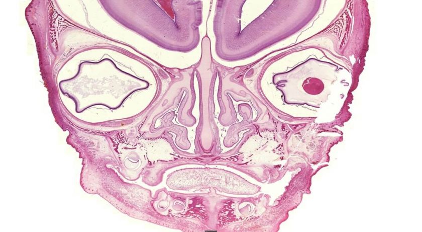
While this may look like a face with two eyes, a nose, and a mouth, this image is actually developing bone captured under the microscope!
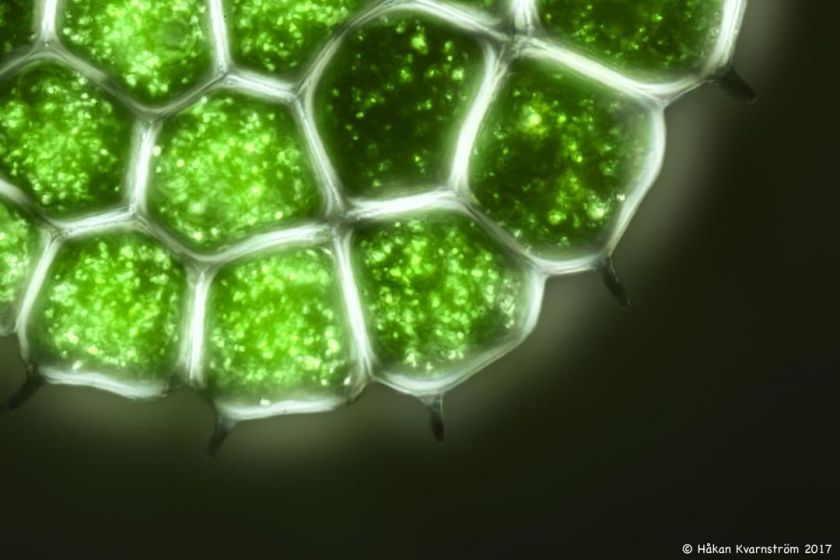
Pediastrum, a genus of green algae, is in the family Hydrodictyaceae. It is a photoautotrophic, nonmotile green algae that inhabits freshwater environments. Pediastrum colonies can contain anywhere from 2 to 128 cells depending on the species of algae. This image was captured in crossed polarized light at approximately 1400x magnification.
Image courtesy of Håkan Kvarnström.
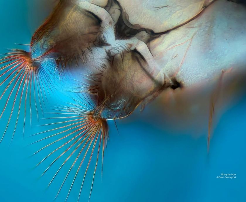
Another image from our European 2018 Image of the Year contest captures the “mouth brushes” of a mosquito. Did you know? Mosquitos remain in the water until they can emerge from the pupal casing as an adult and fly. Captured at 200x magnification on an Olympus BX53 microscope.
Image courtesy of Johann Swanepoel, 2018 Image of the Year Europe Winner.
Bonus image: Sometimes we like to step away from the microscope and have a little fun!
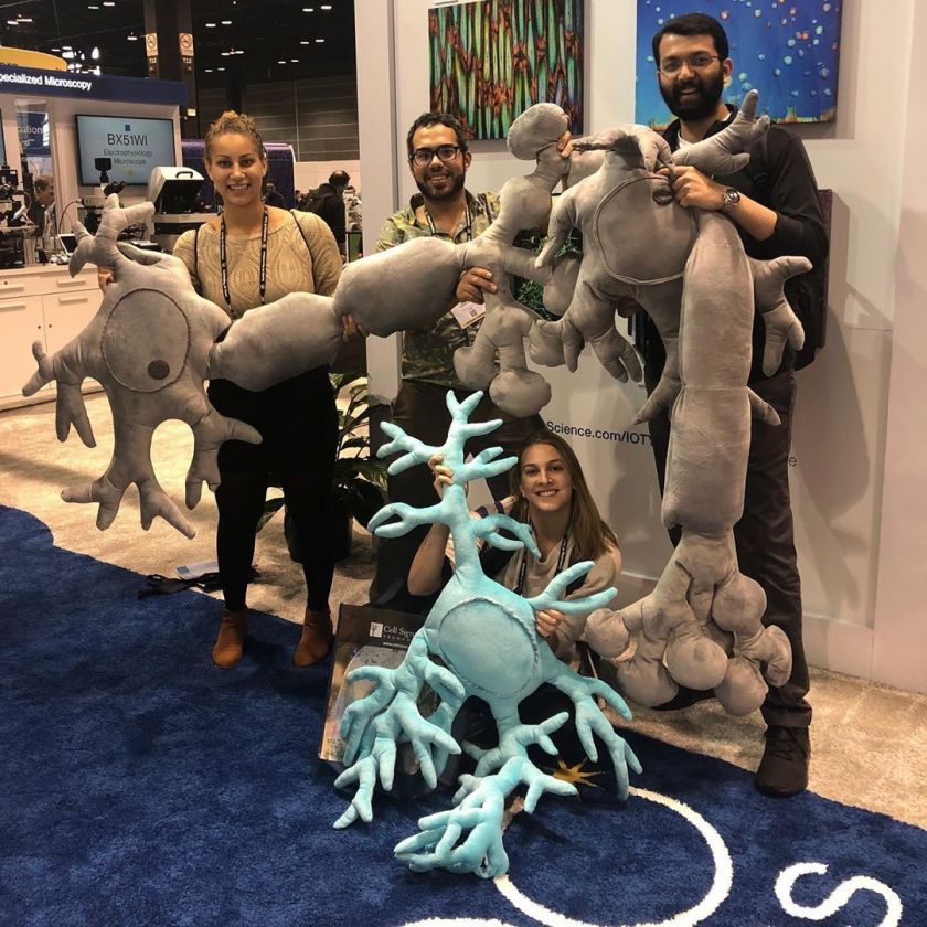
Last week, Olympus Life Science attended the Society for Neuroscience conference in Chicago and brought along a few brainy friends, Natasha the Neuron and Astro the Astrocyte. Thanks to this group for stopping by our booth and striking this synaptic pose!
To see more images like these, be sure to follow us on Instagram at @olympuslifescience!
Interested in sharing your own images?
Visit our image submission site or enter them into our 2019 Global Image of the Year contest.
Related Content
2018 Image of the Year Award Spotlight: 3rd Prize Winner Johann Swanepoel
How to Acquire Microscopy Images that Mimic Human Eye Color Perception
How to Choose the Right Microscope Objective: 10 Questions to Ask
