
Silicone Immersion ObjectivesSilicone immersion objectives are optimized for live cell and live tissue imaging. With a refractive index similar to that of living tissue, silicone immersion objectives offer bright images of cleared tissue. And because silicone oil doesn’t dry out at 37 °C (98.6 °F), time−lapse observations are more reliable and less complex. | 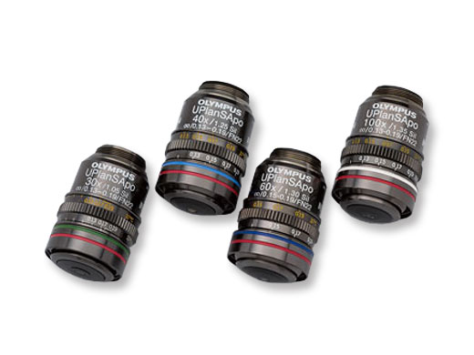 |
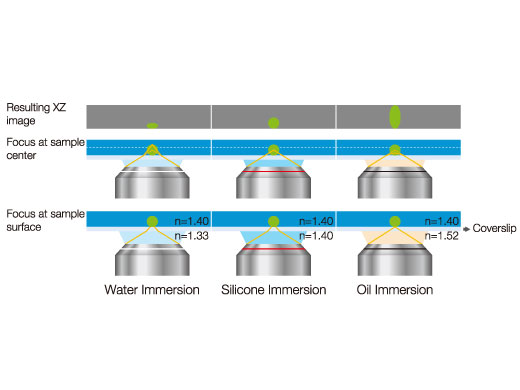 | Effects of Refractive Index Mismatch on Sample ShapeMatching the refractive index of a sample and immersion media is very important to get accurate 3D images. |
|---|
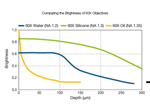
The Brightness of Water/Silicone/Oil 60X ObjectivesOil immersion objectives are the brightest at superficial depths while silicone immersion objectives are brighter than water and oil immersion objectives at all other focus depths. |
|
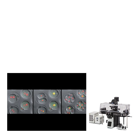
Related Videos | Long–Term, Time–Lapse Imaging of a Live Mouse Embryo (UPLSAPO60XS2)Long-term, time-lapse images of a live mouse embryo captured using a silicone immersion objective (UPLSAPO60XS2). Image data courtesy of Kazuo Yamagata Ph.D., Faculty of Biology–Oriented Science and Technology, Kinki University Reference Stem: Cell Reports. 2014 Jun 3; 2 (6): 910–924. |
|---|
Long–Term, Live–Cell Imaging of Arabidopsis Zygote Embryogenesis (UPLSAPO30XS)The UPLSAPO30XS objective has an NA of 1.05 for brighter images with a higher resolution. The video shows the process of embryogenesis from the early embryo (4–cell stage) to the late embryo recorded over 60 hours. Image data courtesy of Daisuke Kurihara, Ph.D., Optical Technology Group, ERATO Higashiyama Live–Holonics Project, Nagoya University | Related Videos |
Silicone Immersion Objectives Selection Guide
|
Working Distance
(mm) | Magnification | Objective Field Number* | Numerical Aperture | Immersion | Applications | |
|---|---|---|---|---|---|---|
| UPLSAPO100XS | 0.2 | 100X | 22 | 1.35 | Silicone oil | High-resolution for subcellular imaging |
| UPLSAPO60XS2 | 0.3 | 60X | 22 | 1.30 | Silicone oil | High-resolution and long-term, time-lapse imaging of single cells |
| UPLSAPO40XS | 0.3 | 40X | 22 | 1.25 | Silicone oil | Multiple cell imaging with a wider field of view |
| UPLSAPO30XS | 0.8 | 30X | 22 | 1.05 | Silicone oil | Deeper tissue imaging with a wider field of view |
*Maximum field number observable through eyepiece.
Related Products
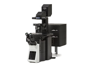
FV3000
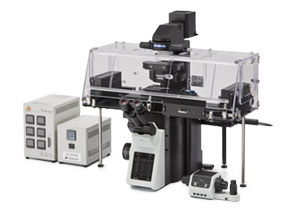
| IXplore Live
| IXplore Spin
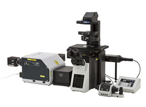
| IXplore SpinSR
|
*Banner Image: By courtesy of Dr. Kazuo Yamagata, Dr. Mayuko Hori, Dr. Tatsuma Yao, Research Institute for Microbial Disease, Osaka University
Sorry, this page is not
available in your country.
Sorry, this page is not
available in your country.