Confocal Microscope Objectives
For any conventional optical microscope configuration, the objective is the most critical component of the system in determining the information content of the image. The contrast and resolution of fine specimen detail, the depth within the specimen from which information can be obtained, and the lateral extent of the image field are all determined by the design of the objective and its performance under the specific conditions employed for the observation. Additional demands are imposed on the objective in scanning confocal techniques, in which this crucial imaging component also serves as the illumination condenser and is often required to perform with high precision at a wide range of wavelengths and at very low light levels without introducing unacceptable image-degrading noise.
Regardless of the function of any other system component, no information can be added to the image that was not initially captured by the objective. Certain intermediate components in the imaging path may perform corrective functions, but their primary performance requirement is that they degrade the fundamental image information received from the objective as little as possible. Traditionally, the primary variables considered in objective selection for a particular application have been the magnification, whether a dry or immersion design is required, and the numerical aperture of the lens system. The development of new imaging techniques driven by technological improvements in lasers, fluorescent dyes, and new specimen labeling capabilities, as well as continuous optical improvements by the microscope manufacturers, have dramatically enhanced progress in certain research areas. This is particularly true in the fields of cell and molecular biology and neuroscience, and the confocal microscope combined with fluorescence techniques has come to be widely relied upon as a research tool.
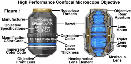
Presented in Figure 1 is a high-performance objective designed specifically for use with laser illumination from violet to near-infrared regions of the spectrum. The objective is a 60X flat-field (plan) apochromat oil immersion model, which is corrected for wavelengths ranging from 405 to 1,000 nanometers. Among the useful applications for this objective is simultaneous fluorescence and differential interference contrast (DIC) observations.
The general demands placed upon the objective in the confocal instrument are similar to those in other critical microscopy applications, although the ever-expanding requirements imposed by new techniques have made it more common than ever to approach the performance limits of these lens systems. The specific limitations of certain applications have made it apparent that other objective characteristics may be as important, or more important, than those traditionally considered paramount. In order to meet essential requirements of currently promising techniques, manufacturers have introduced new optics specifically aimed at optimizing performance for these methods. The development of high numerical aperture, highly corrected silicone oil immersion objectives is an example of response to the dramatic increase in living cell and tissue research, which is by necessity conducted in aqueous media. It is possible that specific requirements in confocal microscopy will lead to the development of optics that less stringently correct one or more of the resolution-compromising aberrations in favor of achieving other design goals that are more critical in confocal performance.
Laser Scanning Microscope Resolution
Resolution in optical systems is commonly accepted to be the minimum separation distance between two specimen features that enables them to be distinguished as separate in the final image. Defined on the basis of a specified contrast required for visual recognition, resolution can be quantified as a function of the light wavelength (λ) and the numerical aperture (NA) of the optical system. Interference between diffracted wavefronts through the len aperture produces the intensity distribution of the Airy disk in the image plane, the diameter of which (d) is given by the following equation, in the ideal diffraction-limited system:
dAiry= 1.22 • λ/NA
The well-known Rayleigh criterion considers two identical points to be barely discernible when they are separated by half the diameter of the Airy disk. Consequently, the Airy radius (r) is equivalent to the lateral resolution, and is defined by modifying the preceding equation. When epifluorescence is utilized, the objective functions as both condenser and objective, and therefore in the following expression, NA represents the objective numerical aperture:
rAiry= 1.22 • λ/(2 • NA)
Diffraction limitations that result in lateral redistribution of intensity from point objects into Airy patterns also produce defocusing along the optical axis, which changes the size and shape of the image point. The three-dimensional intensity point spread function describes the intensity distribution and represents the performance of the optical system in transferring specimen information to the image plane. A range of optical aberrations are characteristic of refractive lens systems, and any that are not corrected by the objective design or compensated by another optical component will alter the intensity distribution that represents each specimen point in the image, degrading the image with respect to the ideal diffraction-limited performance. The relative importance of the potential aberrations in confocal applications must be taken into consideration in selecting objectives for the technique. The principal factors in objective design and performance, as they pertain specifically to the confocal microscope, are discussed in the following sections.
The most widely used confocal microscope configuration for biological applications employs a computer-controlled scanning system to deflect an illuminating laser beam, focused through the objective, across a stationary specimen. This is referred to as off-axis confocal scanning because the scanned beam is deflected to each side of the optical axis and utilizes peripheral regions of the objective lens elements. Some early implementations of scanning techniques in the optical microscope utilized on-axis scanning in which the laser beam remained fixed on the optical axis and the specimen stage or the objective was scanned.
Although in principle the two scanning methods can produce similar results, the requirements with regard to choosing an objective for optimum performance are significantly different for on-axis and off-axis illumination scanning, and the considerations for collection of emitted fluorescence through the objective differ as well. A third scanner type utilizes a spinning Nipkow disk to scan numerous illumination points over the specimen without moving the light source or microscope stage. Nipkow systems are popular in live cell investigations, however, because of their ability to minimize cell damage compared to configurations employing high-intensity scanned laser sources.
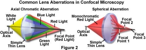
Lens aberrations that affect objective performance may be divided into two categories, including those that are non-chromatic (invariable with wavelength), and the chromatic aberrations, which are wavelength dependent. The chromatic aberrations are characterized as either lateral chromatic aberration or longitudinal chromatic aberration, and the wavelength independent group includes spherical aberration, coma, astigmatism, field curvature, and distortion. Chromatic aberrations and spherical aberration affect the entire image field, while the remaining aberrations are more prevalent in off-axis zones. The chromatic and spherical aberrations (see Figure 2) are the most likely to negatively affect confocal performance. In general, chromatic aberrations cannot be modified procedurally and must be dealt with by design of the optical components. Other artifacts, especially spherical aberration, are commonly exacerbated by improper use of the objective or introduction of mismatched optical components, but can be minimized or compensated by following the appropriate techniques or with adjustments to the optical system.
Spherical Aberration
Spherical aberration is the nonchromatic aberration that is most significant to confocal performance, and it is a manifestation of the property of spherical lens components that causes paraxial and peripheral light rays to be focused in successive planes. A varying degree of refraction for ray paths through different lens zones results in blurring of the focused image of a point source, and produces an asymmetrical intensity change above and below the focal plane. In all modern objectives, spherical aberration is corrected to visually imperceptible levels if the specified operating variables for the objective design are exactly satisfied. Unfortunately, several possibilities exist in practice to allow deviation from the optical design criteria of the objective, and to induce spherical aberration. Because spherical aberration can only be adequately corrected for a precisely specified distance relationship between the objective and the specimen and image planes, the artifact can inadvertently be introduced if the tube length specified for the objective is not maintained. This can occur if the objective is used on a microscope having other than the required tube length in a finitely corrected system, or by the introduction of optical elements, such as filters, into the converging beam path in such a system.
Optimum correction of spherical aberration requires careful attention to the imaging media external to the objective, and this is another potential source of degraded performance. The objective characteristics are fixed by design and cannot be expected to accommodate a range of operating conditions (with the exception of adjustments provided by correction collars). Among the factors that can add significant spherical aberration are poor quality immersion oil between the objective and specimen, nonstandard cover glass thickness, the specimen mounting medium, and the specimen itself. Any material that enters between the specimen and front lens surface of the objective becomes a critical component of the imaging system. Adherence to the design requirements of the objective becomes even more critical at higher numerical aperture. Variations in cover glass thickness or in its refractive index can increase spherical aberration, especially with “dry” (non-immersion) objectives. High numerical aperture dry objectives are generally designed for optimum performance with a cover glass of 0.17-millimeter thickness, having the specimen mounted directly beneath and in contact with the glass.
To enable correction of spherical aberration when non-standard cover glass thickness is used, many objectives are equipped with an adjustable correction collar that can be set to a range of thickness settings. The correction collar operates by translating an internal lens group to alter the objective focal length. Even if a cover glass of the correct thickness is used, the presence of a layer of mounting medium between the glass and specimen deviates from the ideal optical situation, and will increase the degree of spherical aberration. The correction collar may also be utilized to minimize spherical aberration induced by refractive index changes of this nature, which in confocal microscopy causes reduction of intensity at the pinhole and reduction of depth discrimination because of axial focus shifts.
Objectives intended for oil immersion are typically optimized for use with a cover glass of 0.17-millimeter thickness having a refractive index of 1.518 at specified wavelengths, and an immersion oil of precisely defined refractive index. By specifying the operating conditions with respect to the cover glass and immersion medium, spherical aberration can be corrected by the objective design for several values of wavelength (depending upon the type of objective). The importance of matching the refractive indices between each material throughout the light path, from the specimen and mounting medium to the objective front lens element, has been historically one of the most problematic imaging criteria, especially in attempts to achieve high resolution with biological specimens. The refractive index of subcellular components is considerably lower than that of conventional immersion media, and in many cases these refractive indices are uncertain and vary throughout the specimen. Even in fixed materials, the mounting medium refractive index is typically not identical to that of available immersion oils.
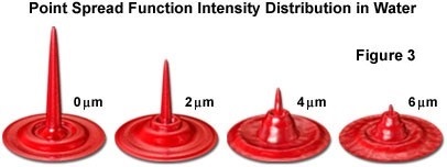
For investigations of dynamic processes in living cells, which are cultured and maintained in physiological saline solution, the mismatch of oil and water refractive indices results in a critical limitation on the performance of oil immersion objectives. The primary problem is that the full potential of the highest numerical aperture oil immersion objectives is not achieved due to the mismatch between the refractive index of water (1.33) and that of immersion oil (approximately 1.5). A critical factor in the application of confocal microscopy is improved fluorescence imaging and three-dimensional representation of thick specimens, and the spherical aberration introduced when oil immersion objectives are used with aqueous specimens limits the depth into the medium at which acceptable images can be obtained. In general, high numerical aperture oil immersion objectives are designed to be used at image planes no more than 15 to 20 micrometers beneath the cover glass. When applied to aqueous specimens, however, the spherical aberration induced at the water-cover glass interface can reach substantial levels at depths as low as 10 micrometers. As an example, the point spread functions illustrated in Figure 3 demonstrate the increasing degree of spherical aberration occurring with a plan apochromat oil immersion objective at penetration depths ranging from zero to eight micrometers.
The interest in utilizing confocal fluorescence techniques for collecting three-dimensional data from aqueous biological specimens was a primary incentive for microscope manufacturers to introduce a number of high numerical aperture, highly corrected water immersion objectives. But water has two disadvantages: it dries up easily so is impractical for long term time-lapse imaging, and it has a low refractive index of 1.33 so cannot be used in the design of high numerical aperture objectives. Thus, manufacturers introduced silicone oil immersion objective for this purpose, because its 1.4 refractive index is close to that of live cells and it does not dry up. Spherical aberration is a major optical limitation in confocal studies of living cells when oil immersion objectives are employed, and the effects increase in proportion to the depth of observation into aqueous media and variable sub-cellular components (see Figure 3). The negative effects on imaging include loss of contrast and signal intensity due to a reduction in the fraction of emitted fluorescence intensity reaching the detector pinhole, loss of resolution of minute specimen features, and reduction of z-axis position accuracy, which affects the integrity of three-dimensional image reconstructions. By utilizing silicone oil immersion objectives when imaging specimens in aqueous media at significant depth beneath the cover glass, the spherical aberration degradation can be avoided or minimized, enabling the full benefits of the confocal technique to be realized. Silicone oil immersion objectives having long working distances are capable of collecting accurate three-dimensional data at depths of more than 200 micrometers in aqueous media.
An oil immersion objective, regardless of the degree of optical correction, is not the optimum choice for imaging a water-immersed specimen. Optimum image quality is possible with this combination only for specimen regions that are in direct contact with the cover glass. At greater depth, the effects of spherical aberration degrade the contrast and resolution and reduce image brightness to the extent that confocal signal-to-noise ratio is drastically reduced. The optimal solution for minimizing induced spherical aberration when imaging at large distances into aqueous media is to use a highly corrected silicone oil immersion objective.
One tactic used to correct spherical aberration is to implement correction collars on water and silicone oil immersion objectives. These adjustment collars provide variable correction for spherical aberration, such as that induced by fluctuations in cover glass thickness. This type of correction collar is also useful in compensating for refractive index differences in physiological media and in cell and tissue constituents, and for variation of refractive index with temperature or solute concentration changes. Because of the variety of factors that can contribute to induced spherical aberration, adjustable correction is a desirable feature of an objective for confocal applications even when a cover glass of optimum thickness is employed.
Off-Axis Optical Aberrations
The common optical aberration referred to as coma primarily affects point-like sources away from the optical axis, producing a streak-like radial distortion of image points, which increases in severity with field angle (see Figure 4). Coma is somewhat similar to spherical aberration in that both are potentially induced by the same factors. This aberration is generally well corrected in modern optical systems by the utilization of appropriate lens elements, and objectives in which coma and spherical aberration are eliminated are classified as aplanatic. Because coma is primarily an off-axis aberration, it has significance to confocal laser scanning systems, which commonly utilize off-axis beam paths, but is not a factor with specimen-scanning (on-axis) confocal microscopes.
Several additional types of geometric aberration are potentially significant to confocal objective performance, and because all are more evident in regions of the image field away from the center, they are more likely to affect performance in laser scanning confocal systems. The optical demands of confocal imaging are such that only highly corrected optics are likely to perform adequately, and significant geometric aberrations will typically have been minimized in objectives from this category. It is beneficial, however, to be aware of these aberrations and the potential problems that they might introduce when objectives are chosen, especially if specific performance characteristics are sacrificed in order to optimize another variable that might be more important in a particular application.
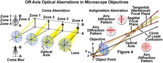
Uncorrected astigmatism can reduce image intensity, sharpness, and contrast, with increasing effects at greater distance from the optical axis (Figure 4). The geometry of an astigmatic image point may be defined by considering two orthogonal planes that are cross sections through the image wavefront. The planes (tangential and sagittal) in an astigmatic system may have different focal distances and exhibit separate radii for a perfectly symmetrical specimen point. This aberration results in symmetrical point-like features that are located off-axis being either radially or tangentially elongated in the image plane, depending upon focus. When the best focus is chosen at a compromise position between the two extremes, the resulting Airy disk is nonsymmetrical, resulting in image degradation. Astigmatism can result from poorly centered elements in a low-quality or damaged objective, and is increased by other misalignments in the optical path of the microscope.
A property of objective lenses described as either field curvature or flatness of field is also a major concern of confocal imaging for wider field of view imaging, especially of tissue sections. Simple spherical lenses focus image points from different regions on a flat specimen onto a curved image surface, which reflects the shape of the lens surface. A flat image plane does not conform to the curved plane of focus, with the result that central and peripheral areas of the field cannot be simultaneously brought into sharp focus. More complex lens designs, consisting of multiple groups of lens elements, are necessary to correct this field curvature and to expand the size of the central zone of sharp focus. Objectives designated as flatfield or plan are corrected optically to produce a wide usable field having sharpness from the center to the edge, with minimal field curvature, at the intermediate image plane. The flatness of the field at the final image plane is also dependent upon intermediate optical elements, including the microscope eyepieces.
Nonlinearity of magnification from the center to the edges of the image field produces geometric distortion of specimen features, causing their true dimensional profiles to be skewed in the image. If present, this effect is readily observed when orthogonal sets of crossed lines are imaged, and rather than appearing straight over the entire image field, the lines are either curved outward or inward at regions away from the field center. These two types of distortion are commonly referred to as barrel and pincushion distortion, respectively. As is the case for field curvature, small amounts of geometric distortion are generally not highly significant in biological applications, but can be crucial in materials science investigations if defect analysis or precise measurements are intended.
Chromatic Aberrations
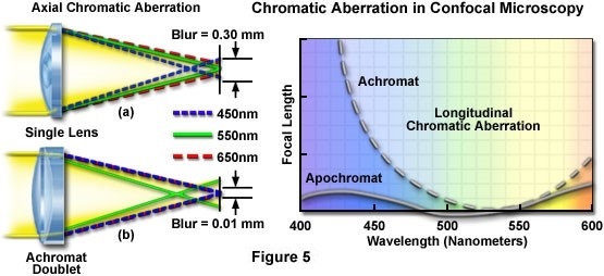
Chromatic aberrations are caused by two fundamental optical phenomena that exhibit wavelength dependence and produce different types of image defect. One type of chromatic aberration is caused by the fact that the refractive indices for all optical glasses vary with wavelength, while the second results from variation in magnification with wavelength. The dependence of refractive index on wavelength (typically referred to as dispersion) produces an effective focal-length difference for light of varying wavelengths. Consequently, for a simple lens consisting of a single glass composition, only one wavelength (or a narrow range of wavelengths) can be precisely focused at the image plane with a particular focus setting. Other wavelengths will be focused either closer or farther from the lens. The resulting spectral dispersion along the optical axis is termed longitudinal chromatic aberration, or alternatively, axial chromatic aberration (see Figure 5). For a point-like source imaged on the optical axis, the gradation in refractive indices cause blue light to be focused nearest the lens, with longer wavelengths converging at focal points progressively farther from the lens. The aberration, if uncorrected, can be visualized as varying color fringes when the image is defocused to either side of the best visual focus.
Uncorrected longitudinal chromatic aberration in an objective used for confocal microscopy can have profound effects, particularly when imaging two or more fluorophores, which depend upon the capability to demonstrate colocalization of fluorescence emission at several wavelengths. When multiple fluorophores are employed, longitudinal chromatic aberration causes the various laser excitation beams to be focused at different points in the specimen, and similarly results in collection of the different emission wavelengths from noncoincident points. This aberration negates the possibility of establishing accurate positions of fluorophores in the z-direction for generation of specimen data in three dimensions.
In scanning confocal techniques, evaluation of the effect of chromatic aberrations on the integrity of images for a particular objective requires consideration of all factors that determine the signal energy that passes through the pinhole and is recorded by the detector. These factors include the excitation laser emission spectrum, the emission peak and bandwidth of each fluorophore, and the spectral sensitivity of the detector. The correction of longitudinal chromatic aberration is usually accomplished in the objective design by the combination of multiple lens elements having different optical characteristics, and the degree of correction forms part of the basis for the classification of objectives into different categories (as discussed below).
The change of lens focal length with wavelength, which produces the axial dispersion characterizing longitudinal chromatic aberration, is also responsible for the occurrence of lateral chromatic aberration (see Figure 5). Because magnification is inversely proportional to focal length, the variation of focal length with wavelength produces a corresponding dependence of magnification on wavelength. If this aberration is not corrected in the objective, sharp edges in the image may be surrounded by red or blue color fringes. The magnification of the blue wavelength component from the signal may vary from that of the red component by approximately 1 to 2 percent in uncorrected objectives. When present in a confocal scanning optical system, lateral chromatic aberration can result in a loss of signal at the pinhole, because of the possibility that emitted light will be imaged at a location that is either nearer to or farther from the optical axis than its true specimen location, depending upon the spectral composition of the light.
Because the two types of chromatic aberration are related, objectives that are highly corrected for longitudinal chromatic aberration generally exhibit minimal lateral chromatic aberration as well. Different approaches are taken by microscope manufacturers in correction of the various factors affecting optical performance, and careful matching of system components is essential to obtaining the maximum correction of aberrations. In confocal fluorescence microscopy, uncorrected lateral chromatic aberration results in similar problems to those of the longitudinal variety in that errors are introduced in mapping the locations of fluorophores emitting at different wavelengths.
For much of the history of optical microscope manufacture, a standard for objective design was followed that required the objective to form a real image at a specified fixed distance from the mounting surface of the objective, corresponding to the front focal plane of the eyepiece. Configurations based on the direct formation of a real intermediate image by the objective are referred to as finite systems, and the distance to the intermediate image is referred to as the tube length of the objective. Although at one time the various manufacturers standardized their finite optical systems on the same tube length and parfocal distance, different approaches have been employed to compensate for lens aberrations, and this factor must be considered if optical components from different manufacturers are used in combination. Finite systems may be designed to provide full aberration correction in the objective, or a known residual level of lateral chromatic aberration may be allowed in the objective-formed image, which is compensated by the eyepiece.
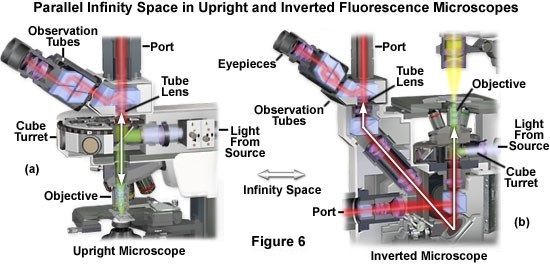
In the infinity optical system, light rays leaving the objective are not focused, but remain parallel until being converged at the intermediate image plane by a tube lens (occasionally referred to as a telan lens). Most major microscope manufacturers have developed infinity-corrected optical systems, which have the intrinsic benefit that light rays from the objective, focused to infinity, are relatively insensitive to additional optical components placed in the “infinity space” between the objective and tube lens. Although the tube lens can perform some correction of residual aberrations, there are advantages in versatility of designs that provide full correction within the objective, with the tube lens being optically neutral. Presented in Figure 6 are the infinity light pathways for typical upright and inverted fluorescence microscopes. The double-headed white arrow in each microscope diagram indicates the parallel light path between the objective rear aperture and the tube lens.
Different manufacturers adhere to different design specifications in their infinity systems, including tube lens focal length and length of the infinity space. Infinity-corrected optical systems enable the objective to be moved for focusing instead of the microscope stage, a distinct advantage in applications requiring delicate manipulation of the specimen during observation. However, the principal practical benefit of the infinity space is that auxiliary optical components, such as polarized light analyzers, filters, and differential interference contrast (DIC) prisms, can be added without major concern for their refractive properties or thickness. As long as they have plane parallel surfaces, the added elements have negligible effect on image quality. In contrast, optical components cannot be placed into the beam path of a finitely corrected system, which is converging between the objective and intermediate image plane, without introducing image shift and other aberrations that vary with the thickness and refractive index of the added elements.
Optical Correction in Microscope Objectives
Regardless of whether a finite or infinity-corrected optical system is employed, the combined performance criteria of the objectives designed for the system must be taken into account in determining which objectives will meet the requirements of a particular imaging technique. Objectives are typically grouped into performance categories based upon their degree of optical correction. As discussed, the relative importance of various aberrations and the performance criteria they affect depend upon the manner in which the objective is used and the essential requirements of the application. The most general grouping of objectives is typically based on their degree of chromatic aberration correction, although the traditional objective categories have become much less distinct recently, owing to significant technological advances in optical design and manufacturing. Much of the descriptive nomenclature is still valid and widely used, and an understanding of the terms employed and how they pertain to confocal applications is important when the various objectives available are being evaluated.
Chromatic aberrations are a result of dispersion in the optical glass used to fabricate a lens, and are typically corrected by utilizing a combination of lens elements having different dispersive properties. Traditionally, objectives having the minimal correction for chromatic aberration are categorized as achromats (see Figure 7) and are usually constructed using glass with “normal” dispersion. Glasses of this type exhibit a nearly linear decrease of refractive index with increasing wavelength, and include crown and flint glasses. Crown glass usually exhibits low refractive index and low dispersion, in contrast to flint glass, which typically has high refractive index and high dispersion. In older achromatic objectives, two or more lens elements of these glass types were combined to provide chromatic aberration correction that brings red and blue light to a common focus, and also corrects spherical aberration for green light. Modern achromatic objectives typically have additional correction for spherical aberration, and significant correction for field curvature. If the objective is corrected to extend flatness of field over the entire image field, it is given the designation plan achromat. In general, achromatic objectives are suitable for conventional brightfield visual observation, and for photomicrography and digital imaging with the additional (plan) correction for field curvature.
In order for the objective to achieve improved chromatic aberration correction, glass types having abnormal dispersion over a portion of the spectrum are required. In glasses that exhibit nonlinear change of refractive index with wavelength in the red or blue spectral region, the chromatic aberrations can be offset to enable simultaneous focus of more than two wavelengths. Crystalline fluorite was one of the first materials found to provide suitable optical properties in reducing the uncorrected secondary spectrum responsible for the green or purple fringes bordering sharp edges, which characterize images acquired with achromats. In combination with various optical glasses, fluorite elements provide the capability to correct chromatic lens aberrations for three wavelengths (colors) and spherical aberration for two. Additionally, such highly corrected objectives have improved transmission characteristics in the ultraviolet spectral region.
More recent technological developments have provided new optical glass formulations and lens shaping capabilities that produce dispersion properties similar to those of fluorite elements, and most manufacturers produce a line of fluor objectives that have optical corrections approaching those of the highest category (Figure 7). These objectives are sometimes referred to as semi-apochromats, and may or may not contain fluorite elements, but because of their optical characteristics, are nevertheless designated by different manufacturers with names such as Fl and PlanFl. Current objectives in this category are available in many configurations, including models for use with multiple immersion media, and are suitable for use in brightfield, fluorescence, phase contrast, polarized light, and differential interference contrast, as well as some confocal and multiphoton applications.
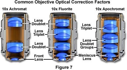
Objectives classified as apochromats are generally those that have the highest level of correction for chromatic and spherical aberrations (see Figure 7). These objectives typically have the highest available numerical apertures for a given magnification, and both chromatic and spherical aberrations are corrected for at least three wavelengths. With nearly complete correction of aberrations, apochromatic objectives are generally suitable for any microscopy technique, although all of the specific performance requirements for the technique being employed must be considered. In spite of the exceptional degree of optical correction exhibited by apochromats, because they utilize triplet or doublet lens elements, they sometimes sacrifice other important specifications, such as numerical aperture or flatness. Newer commercial manufacturing techniques have overcome this compromise through a novel lens polishing technology enabling ultrathin lens elements. By combining ultrathin convex and concave lenses, these objectives achieve chromatic aberration correction and higher transmittance at spectral range from violet to NIR, higher numerical aperture, and expanded flatness.
Confocal fluorescence applications can be severely limited if the objective aberrations are not similarly corrected for both excitation and emission wavelengths, a requirement made more difficult to satisfy when multiple fluorophores are utilized or when the difference between excitation and emission wavelengths is large. In order to achieve maximum detected photon energy, parfocality between the illuminating spot and the detected region imaged at the pinhole must be maintained. Many objectives, even in the apochromat class, do not provide adequate correction for fluorescence techniques combining ultraviolet excitation with visible-region emission. Additional optical components may be employed at the ultraviolet laser source to compensate for the failure of the objective to accommodate the wide range of wavelengths, although this adds expense and considerable operational complexity to the confocal system.
High-performance water immersion objectives and silicone oil immersion objectives are specifically designed to satisfy requirements imposed by live cell imaging research areas. A primary goal is achieving optimum performance in confocal fluorescence microscopy of biological specimens, including live cells and tissue supported in physiological media. It is essential to recognize that regardless of the level of aberration correction incorporated into an objective's design, additional aberrations can be introduced into the optical path outside the objective by violating the design's operating requirements, and these can degrade the performance of the best components.
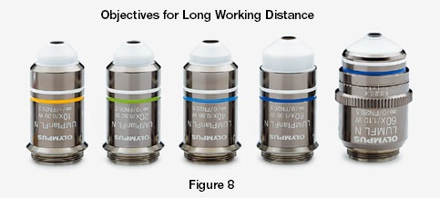
Objectives designed for direct immersion or water dipping, which feature specialized ceramic or polymeric nosecones, are available for live cell and physiological studies. These investigations often require access to the specimen for measurements or other manipulations, and cannot be done with a cover glass in place. Generally, water dipping objectives are in the fluorite category, have high transmission in both the ultraviolet and infrared spectral regions, and are designed with long working distance specifications and narrow profile of the insertion (dipping) portion at the nose of the objective. The smaller angular nose on the objective is intended to enable maximum access to the specimen for attachment of microelectrodes or to perform other operations during observation. Long working distances, which may be several millimeters with some objectives of this style, also offer greater access to the specimen. The immersion portion of many dipping objectives is constructed from ceramic or another inert insulating material for electrical isolation and chemical resistance. Their combination of characteristics makes water dipping objectives suitable for live specimen confocal, multiphoton, and many other imaging methodologies.
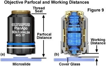
In addition to magnification, numerical aperture, and degree of optical correction, an objective’s working distance has particular significance in confocal microscopy and other digital microscopy techniques used for acquiring three-dimensional specimen information. The working distance, for an objective requiring a cover glass, is defined as the distance between the top surface of the cover glass and the front lens element when a specimen plane in contact with the cover glass is in focus (Figure 9(b)). This distance is important when confocal imaging is performed at various depths in the specimen because it determines the maximum focal penetration depth that is attainable before the objective contacts the top of the cover glass. The working distance, therefore, limits the range along the z-axis from which specimen data can be collected.
As an example, an objective with 0.20-millimeter (200 micrometers) working distance can be focused to a maximum depth of 200 micrometers below the cover glass before the objective contacts the cover glass upper surface. Within limits, the working distance can be varied by optical design, although it generally decreases as magnification, numerical aperture, and optical correction is increased. Maintaining large numerical aperture and a high degree of aberration correction with long working distance is limited by simple geometric constraints imposed by the shape of the lens elements and the number required in a highly corrected objective. The specifications of many currently available objectives represent remarkable achievements in performance realized by the availability of improved optical glasses, lens coatings, and computer design capabilities.
The fact that signal intensity is proportional to the square of the numerical aperture has implications in the choice of optimum objectives for confocal microscopy, in some cases altering the relative importance of various performance factors. When utilizing scanning confocal systems with a scan zoom feature, it is seldom necessary to employ 100x optical magnification, and high numerical aperture and high corrected 40X and 60X objectives may be more suitable. They can acquire wide field of view images using a lower scan zoom and high-resolution subcellular structure images using a higher scan zoom.
Many popular techniques, such as immunofluorescence imaging, inherently suffer from such low light levels that the transmission properties of the optical system are of crucial importance. Only a few photons may be available for the detection of minute specimen details, and as a consequence a relatively minor difference in transmission percentage at the wavelength of fluorophore excitation or emission can be critical in determining the detectability of a labeled feature. In some situations, the objective’s transmission properties may have greater significance than other specifications such as flatness of field and aberration correction, which require additional lens elements and coatings that can increase light loss in a critical wavelength band. However, advances in optical technology avoid this transmittance compromise through unique lens manufacturing technology.
The number of specialized techniques being applied to problems in cellular and molecular biology, as well as in other fields, has resulted in changes to the demands placed on optical imaging systems, particularly with regard to the microscope objective. The dramatic increase in use of confocal scanning techniques for three-dimensional investigations of biological specimens has had a major influence on the products that have been commercially developed, although methods such as laser trapping, multiphoton excitation, fluorescence resonance energy transfer (FRET), fluorescence in situ hybridization (FISH), and infrared differential interference contrast have also introduced new requirements. A number of the parameters for specialized techniques have made it apparent that certain traditional objective performance criteria may be able to be at least partially sacrificed in order to improve other more critical specifications. Dramatic advancements in computer-aided lens design and optical glass formulations, in addition to those in antireflection coating technology, have resulted in the introduction of improved optics that are able to meet many of the new requirements, with few or no trade-offs in conventional wide field microscopy. Examples of these include high-performance silicone oil immersion objectives with higher numerical aperture, increased working distance, spherical aberration correction collars, and objectives with enhanced ultraviolet and infrared transmission. Additionally, objectives manufactured with ultrathin lenses enable researchers to achieve high numerical aperture, high transmittance, wider spectral range chromatic aberration correction, and expanded flatness without compromising on any of these specifications.
Many modern objectives are suitable for confocal microscopy when they are used in accordance with their design specifications. High-resolution examination and accurate three-dimensional imaging of thick aqueous specimens with a laser scanning confocal microscope is particularly demanding and requires an objective satisfying all of the following criteria: high numerical aperture for efficient fluorescence collection, long working distance enabling maximum penetration depth, flat field for accurate three-dimensional reconstruction, low axial chromatic aberration for parfocality of multiple fluorophores, and low lateral chromatic aberration to enable precise registration of multiple fluorescence images, in addition to high transmission at the excitation and emission wavelengths.
Sorry, this page is not
available in your country.


