Challenges Solutions of Confocal Microscopy in Live Cell Imaging
Duration: 30 minutes
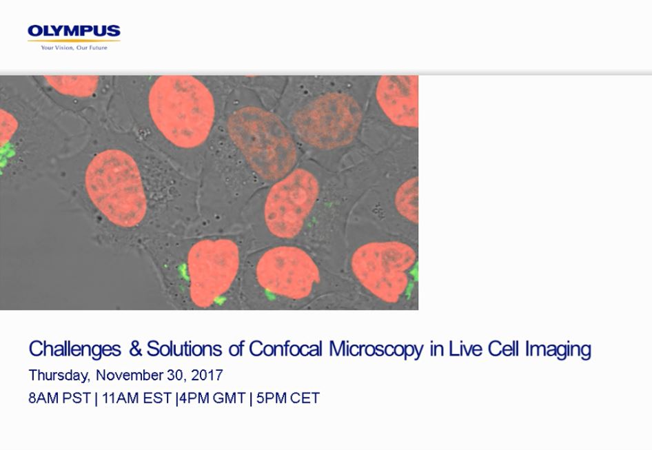
Live cell imaging is a powerful tool to visualize dynamic processes in cells and tissues, ultimately showing changes happening in their biological environment. The need for higher resolution, 3D information, and better contrast often leads to the usage of confocal microscopy.
However, there are different strategies to overcome these effects, including the use of more sensitive detectors or reducing the excitation time per pixel to avoid dark states of fluorophores.
In this shortcast we will discuss the image formation on spinning disk and point scanning confocals and compare them across various applications.
Presenter:
Florian Eich
Senior Business Development Manager Life Science
Olympus Life Science
Challenges Solutions of Confocal Microscopy in Live Cell Imaging
Duration: 30 minutes
Live cell imaging is a powerful tool to visualize dynamic processes in cells and tissues, ultimately showing changes happening in their biological environment. The need for higher resolution, 3D information, and better contrast often leads to the usage of confocal microscopy.
However, there are different strategies to overcome these effects, including the use of more sensitive detectors or reducing the excitation time per pixel to avoid dark states of fluorophores.
In this shortcast we will discuss the image formation on spinning disk and point scanning confocals and compare them across various applications.
Presenter:
Florian Eich
Senior Business Development Manager Life Science
Olympus Life Science
Not Available in Your Country
Sorry, this page is not
available in your country.