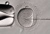
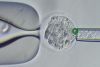
To ensure successful time-lapse observation of live cells, your microscope system must maintain constant environmental control, suppress any phototoxicity and photobleaching, and provide stable imaging. We offer reliable live cell imaging solutions featuring silicone oil immersion objectives optimized to the refractive index of live cells. The live cell imaging software that supports our inverted imaging platforms enables you to quickly and easily set up complex experiment protocols and
configure precise functions such as incubation, maintaining the focus position using the motorized stage, and illumination control. Each inverted live cell imaging platform meets the broad demands of live cell observation applications, from cellular level interactions to small structure in organelles, through the combined capabilities of TIRF, confocal, and super resolution imaging.
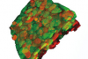
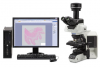
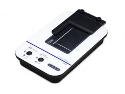
The APEXVIEW APX100 all-in-one microscope makes it fast and simple to acquire expert-quality microscope images. Built with renowned Olympus optics, an intuitive user interface, a powerful AI, and a suite of smart features, the APX100 system combines the ease of use of an all-in-one-microscope with high-quality image data to fit your research needs.
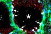
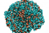
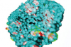
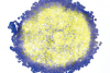
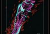
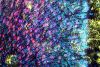
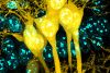
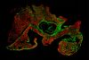
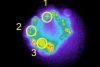
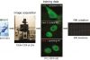
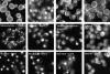
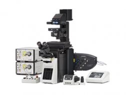
The IXplore SpinSR system is our confocal super resolution microscope optimized for 3D imaging of live cell specimens. Like the IXplore Spin system, it has a spinning disk system for fast 3D imaging while limiting phototoxicity and bleaching. However, it achieves super resolution images down to 120 nm XY and enables you to switch between widefield, confocal, and super resolution with the click of a button.
Sharp, clear super resolution imaging down to 120 nm XY, owing to Olympus Super Resolution (OSR)
Prolonged cell viability in confocal time-lapse imaging due to less phototoxicity and bleaching
Use two cameras simultaneously to achieve fast, two-color super-resolution imaging
Super resolution imaging with the world’s first plan apochromat objectives with a numerical aperture (NA) of 1.5*
* As of November 2018. According to Olympus research.
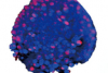
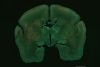
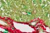
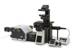
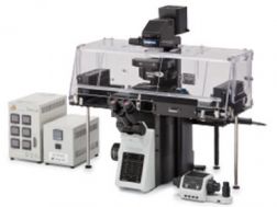
Designed to reduce photobleaching and phototoxicity, the IXplore Live system is optimized for physiological experiments involving live cell and tissue observation. Offering precise environmental control and enhanced rigidity, it supports long-term cell viability and stability for time-lapse imaging applications, such as in cancer, stem cell, and brain research.
Maintain focus accurately and reliably in time-lapse experiments with TruFocus™ Z-drift compensation system
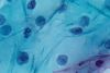
Discover the real morphology of your cells with Olympus silicone immersion optics
Real-time controller helps limit cell disturbance, enabling physiologically relevant data
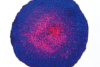
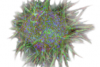
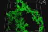
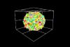
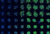
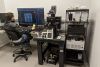
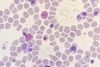
The IXplore Spin system features a spinning disk confocal unit that enables fast 3D image acquisition, a large field of view, and prolonged cell viability in time-lapse experiments. Researchers can use it to perform rapid 3D confocal imaging with high resolution and contrast at greater depths for imaging into thicker samples. The spinning disk also helps to cut down on photobleaching and phototoxicity of samples upon excitation.
Real-time controller (U-RTCE) helps optimize the device’s speed and precision during automated acquisition
TruFocus™ Z-drift compensation system maintains focus for each frame
Precise 3D imaging with improved light collection using X Line™ objectives
Upgrade to the IXplore SpinSR super resolution system as your research progresses
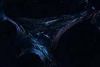
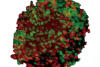
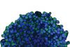
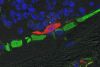
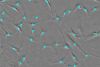
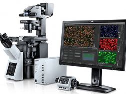
For membrane dynamics, single-molecule detection, and colocalization experiments, the IXplore TIRF system enables sensitive simultaneous multicolor TIRF (total internal reflection fluorescence) imaging for up to four colors. Olympus’ cellTIRF system provides stable motorized individual laser-angle control, providing equal evanescent wave penetration for high-contrast, low noise images. Our TIRF objectives feature high SNR, high NA, and correction collars to adjust for cover glass thicknesses and temperature.
Exact colocalization of up to four markers thanks to individual penetration depth control
Take advantage of Olympus’ TIRF objective with the world's highest NA of 1.7*
Intuitive setup of complex experiments with the Graphical Experiment Manager (GEM), cellFRAP, and U-RTCE
* As of July 25, 2017. According to Olympus research.
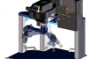
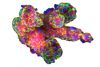
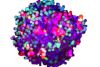
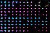
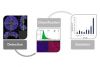
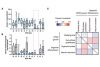
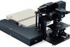
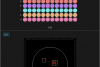
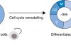
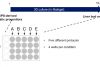
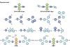
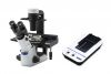
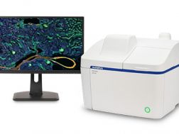
Achieve fully automated image acquisition and data analysis of biological samples using the scanR high-content screening station. Design individualized assays for cell cycle, protein localiazation, intracellular transport and more. Modular hardware is compatible with a range of additional systems, including spinning disk confocal, robot loading, incubation, TIRF, and FRAP systems.
Fast and precise image acquisition and analysis
Image cytometry based approach enables easy and detailed visualization of results
Expand your capabilities with modules such as self-learning AI, kinetic parameter measuring, high-speed 3D deconvolution, and more
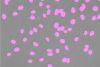
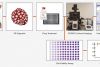
Remotely monitor, analyze, and share your cell cultures’ health, cell count, and confluency using the reliable quantitative data provided by the automated CM30 incubation monitoring system. The system enables label-free observation, reduces the risk of damage to your cultures, and standardizes your culture workflow.
Automatically collects quantitative data on the health and confluency of your cultures
Monitor, analyze, and share your cultures' progress remotely from a PC or tablet
Equipped with oblique epi-illumination for label-free observation
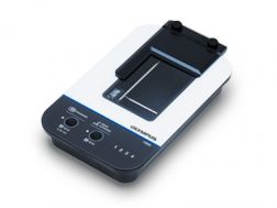
Remotely monitor, analyze, and share your cell cultures’ health, cell count, and confluency using the reliable quantitative data provided by the automated CM20 incubation monitoring system. The system enables label-free observation, reduces the risk of damage to your cultures, and standardizes your culture workflow.
Automatically collects quantitative data on the health and confluency of your cultures
Monitor, analyze, and share your cultures' progress remotely from a PC or tablet
Equipped with oblique epi-illumination for label-free observation