Evident LogoOlympus Logo
High-Content Imaging and Analysis Solutions

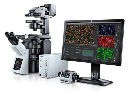
Your discovery research is constantly evolving, and the right tools are critical to an efficient workflow. We provide a range of imaging system solutions with the optical performance you need to make the most of our powerful software tools, from artificial intelligence powered analysis with TruAI technology to true 3D analysis using our NoviSight software.

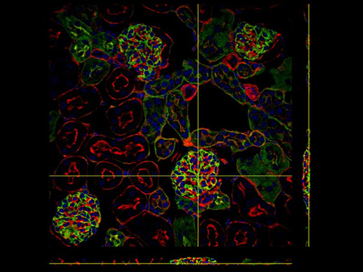
With the advance of more physiologically relevant 3D models, confocal and multiphoton imaging solutions equipped with the right optics provide better images for better results. Coupled with our NoviSight 3D software for high-content analysis (HCA), our high-quality microscope systems can resolve your most challenging samples by revolutionizing traditional 2D screening methods with TruAI deep learning and providing true 3D analysis.
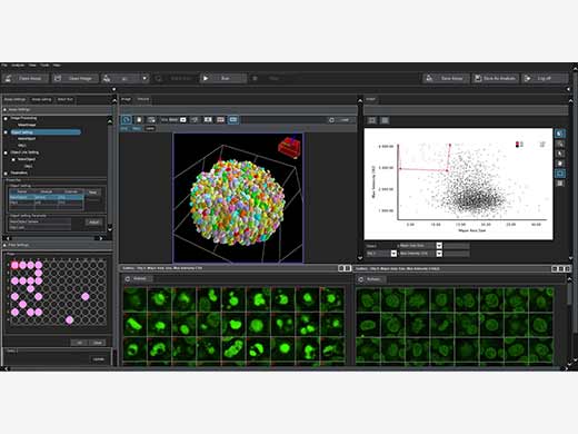
NoviSight Software

Novisight

3D High-Content Analysis

  • Obtain true object counts and morphological measurements in 3D
  • Statistical reporting of microplate data
  • Dynamically linked data, from image acquisition to analysis
  • Standard assays and algorithm customization service

Learn more 

ScanR High-Content Screening

  • Powered by TruAI deep-learning analysis
  • Smooth workflow for fast results
  • Robust, modular hardware
  • Ideal for assay development
  • Deep imaging application support and consultation

Learn more 

ScanR High-Content Screening

FV3000RS High-Speed Scanning Microscope

FV3000RS Microscope

Faster and Deeper Confocal Imaging with High Resolution

  • Superior optical sectioning at depth, compared to fixed aperture systems, through the use of a single, adjustable confocal aperture
  • Simultaneous multichannel spectral detection
  • Macro-to-micro functionality
  • Advanced optical performance deep in live spheroids and tissues
  • Range of customized solutions

Learn more 

FVMPE-RS Microscope

Multiphoton Imaging for Thick Samples

  • Image whole organisms, tissues, tumoroids
  • Imaging in live and fixed tissues
  • In vivo imaging
  • Microplate compatible
  • Range of customized solutions

Learn more 

FVMPE-RS

IXplore Spin Microscope System

IXplore Spin

Confocal Imaging of Rapid Cell Dynamics

  • Ultrafast spinning disk confocal with the Yokogawa CSU-W1 scanner unit
  • Precise 3D imaging with improved depth performance using silicone oil immersion objectives
  • Multi-point illumination for reduced phototoxicity during time lapse experiments
  • Expandable with modular upgrades for super-resolution, TIRF, and FRAP

Learn more 

IXplore SpinSR Microscope System

Olympus Super Resolution Imaging for Live Cell Samples

  • Olympus Super Resolution (OSR) imaging down to 120 nm XY resolution
  • Enables easier quantification of subcellular structures, such as lipid rafts, membrane clusters, and vesicles
  • Compatible with standard dyes and fluorophores—no special SR reagents required
  • Suitable for live cell imaging in multiwell vessels and microplates with recommended lenses

Learn more 

IXplore SpinSR


Sorry, this page is not
available in your country.

Sorry, this page is not
available in your country.

Sorry, this page is not available in your country