
Many researchers and lab workers require microscopy images to show the exact color they see through their eyepieces. For instance, pathologists use colors to identify tissues, while cytologists identify cell type based on color difference with a Papanicolaou stain.
So, how do you reproduce human eye color perception in your microscope images? To do so, you must first consider how your own eyes perceive color compared to microscope cameras and monitors.
Today’s blog post will explore how human color perception works and reveal how Olympus’ camera technology can replicate it for microscopy.
History of the Human Eyes – Why We Can’t See Color at Night
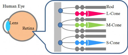 | A human eye has two different types of cells: rod cells and cone cells. Rod cells are more sensitive than cone cells, allowing us to detect dim light and see at night. Humans have about 20x more rod cells than cone cells, perhaps because our ancestors spent time awake in the dark. But since these cells cannot distinguish colors, our eyes have trouble detecting colors at night. |
Cone cells, on the other hand, cannot detect dim light but can identify colors. To do so, they use three types of cells:
- L cone for longer wavelength light sensing
- M cone for middle wavelength light sensing
- S cone for shorter wavelength sensing
Evolution of Eyesight
Our ancient ancestors originally had four types of cones: ultraviolet (UV), blue, green, and red light. Today, fishes and birds still can see with this wide wavelength range. But after our ancestors lost cones for UV and green, we received the M cone as a mutation of the L cone for green light detection.
This better color resolution might have helped our ancestors gather food in the daylight. Our evolutionary history has led to our current spectrum response, shown in the table below. As you can see, M cones and L cones are close, because the M cone evolved from the L cone.
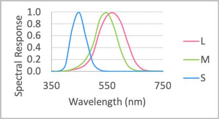
Fig. 1 Spectrum sensitivity of the human eye (sensitivity has been normalized for each peak)
Mimicking Human Eyesight with a Microscope Camera
On the other hand, camera sensors, like a charge-coupled device (CCD) or complementary metal-oxide semiconductor (CMOS) sensor with a Bayer filter, feature a different spectrum sensitivity, as shown in the table below. In addition, since a PC monitor’s light spectrum is based on an RGB signal, it cannot produce the same spectrum as the physical sample. Olympus cameras use a special treatment to reproduce the color you see through your eye on your computer monitor.
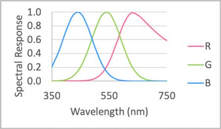
Fig. 2 Spectrum sensitivity of a camera sensor
How Microscope Cameras Deliver Accurate Color Reproduction
Most camera sensors have a sensitivity over 700 nm, which causes images to look reddish from the IR light. Since human eyes cannot see near IR or IR light, our color cameras use an infrared (IR) cut filter. Then we apply a microscopy dedicated image processing technique to convert signals from the sensor to the image data, which can be displayed on a monitor to mimic the response from your cone cells (Fig. 3).
This is easier said than done because you must understand the microscope’s illumination spectrum, as well as the spectrum character of the specimen, dyes, colors, camera, and PC monitor. But with our microscope data and knowledge of samples used commonly for biology, we developed microscopy color reproduction techniques for our microscope cameras.
This technology enables us to offer a wide range of microscope cameras with excellent color reproduction. Use our camera selector to find the best one for your application.
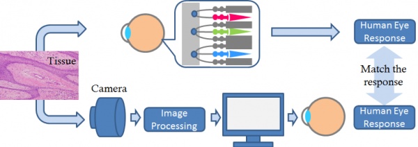
Fig. 3 Olympus’ color reproduction technique to mimic the human eye response
Related Content
Camera Selection: Choose the Best Camera for Your Application
