Automatic Objective Changeover
Automatic Objective Changeover - Java Tutorial
The ergonomic design of stress free microscopes that are easy to operate is a primary goal for all of the manufacturers. This interactive tutorial examines the automatic objective changeover design in clinical microscopes, a feature that is quickly performed with a foot or hand switch to reduce the frequency of repetitive hand motion and ease operator discomfort.
The tutorial initializes with the automatic nosepiece feature of an ergonomically designed modern microscope (the Olympus BX45A) appearing in the window. In order to operate the tutorial, use the Swap Objectives button to toggle between the 10x and 40x objective and simulate how these components are rapidly interchanged through the use of a remote hand or foot switch.
New ergonomic microscope designs incorporate a two-position motorized nosepiece, which eliminates repetitive motion of rotating the nosepiece to interchange objectives. In addition, the 10x objective is often intensity equalized, so that it is not necessary for the operator to adjust the illumination intensity (lamp voltage) when changing between low and higher magnification objectives. The discomfort, and potential damage, that is caused when the eyes are exposed to sudden changes in light intensity is eliminated in this design.
Upright compound clinical microscopes command the second greatest market share (next to stereo microscopes) and are sold by the thousands every year. The design of these microscopes has also received considerable attention from the manufacturers with respect to ergonomic factors. Included in the new list of features are one-handed stage and focus control, optimum eye-level positioning, low-profile stages, and rigid instrument body standards, all contributing to strain-free posture for operators while examining specimens.
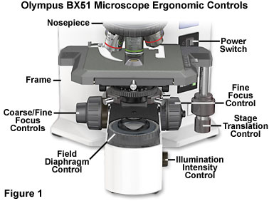
Perhaps the most significant feature of new compound microscope designs is positioning of the stage translation and focus control knobs in an equidistant configuration from the operator (Figure 1). This allows a more relaxed posture with the hands resting comfortably on the desktop. In addition, operators are no longer required to twist their shoulders to simultaneously manipulate the stage and focus controls, considerably reducing the amount of strain associated with long-term observation. Other frequently used microscope controls, such as the field diaphragm, light intensity potentiometer, and auto-illumination photomicrography preset switch, are located in the front of the microscope base at desktop height and within easy reach of the operator (see Figure 1 for details). Push-button filter engagement levers are also positioned near the main controls to further enhance operational ease. Many microscopes are equipped with a refocusing stopper that allows quick change of specimens with an instant return to focus. In addition, coarse focus tension controls allow the operator to customize the action of z-direction stage translation to suit individual preferences. A few microscopes also offer detachable fine focusing knobs that can be fitted to either side of the instrument to suit the operator's preferences, and some knobs are coated to enhance traction and allow easy operation with only a single finger.
Lowered stage assemblies, in recent instrument designs, have heights ranging from approximately five to eight inches, considerably lower than those found on previous microscopes. Many also incorporate space-saving dual pulley or pulley and guide rod systems that replace older rack and pinion mechanisms to govern stage movement. Some models feature focusing adjustments that translate the nosepiece, rather than the entire stage, to maintain a constant stage height, reducing the amount of forearm movement necessary to move the stage or change the specimen. More advanced designs eliminate protrusion of the x-directional guide from the side of the stage, thus reducing interference with focusing action. Rotational movement of modern stages often exceeds 200 degrees and some can even achieve over 250 degrees of rotation about the microscope optical axis. Any microscopist who has spent time framing images for capture on film or by digital imaging will appreciate this advanced feature, which not only saves time, but eliminates a considerable amount of frustration.
对不起,此内容在您的国家不适用。


