
Cell division is an essential process for all living organisms. Prior to each division, the genetic information of the cell, which is stored in the DNA, must be accurately copied. This copying process is known as DNA replication, and its proper regulation plays a vital role in the health and survival of an individual. Understanding how cells regulate DNA replication to achieve high accuracy in genome duplication is one of the fundamental questions in biomedical research. Errors generated during DNA replication can lead to genome instability associated with severe diseases of human health such as cancer. In fact, previous research has estimated that up to two-thirds of all cancers are caused by the accumulation of errors during DNA replication.
Meet a Trailblazer in the Field of DNA Replication
How cells duplicate their DNA is also the research focus of Hana Polasek-Sedlackova, PhD. Hana got an early introduction to the lab and DNA research while participating in a scientific research internship at Masaryk University during secondary school. Since then, she’s been a rising star in the field. Hana’s undergraduate thesis won the renowned global Undergraduate Award in Life Sciences, which is often referred to as the ‘junior Nobel Prize.’ After graduation, she received a scholarship from the Novo Nordisk Foundation to attend the Copenhagen Bioscience PhD program. With this scholarship, Hana joined the laboratory of Professor Jiri Lukas at the Novo Nordisk Foundation Center for Protein Research at the University of Copenhagen as a PhD student and continued her postdoctoral training there. Her research work was published in leading scientific journals, including Science and Nature. Recently, she started her own independent research group at the Institute of Biophysics (IBP) of the Czech Academy of Sciences in Brno, Czech Republic.
One of her contributions to science was solving the MCM-paradox, which has been debated since the 90s among scientists in the field. It described seemingly contradicting results between microscopy and other scientific methods used to elucidate the events during replication. This contradiction has now been successfully resolved with the aid of our scanR high-content screening station, of which Hana is an avid user. We met Hana during a scanR user meeting at the Institute of Molecular Genetics in Prague and took the opportunity to ask her about her experience with the system and how it impacts her research.
Our Interview with Hana Polasek-Sedlackova
Evident: When you started your own research group at the IBP, the scanR high-content screening station was the first microscope you acquired for the group. What made you choose this system??
Hana Polasek-Sedlackova: During my professional experience, I have had the opportunity to work with several microscopes. Based on my observations, I quickly realized that there are three important parameters that one should consider when choosing a microscope. These parameters are hardware, software, and customer service. After carefully considering these criteria, I found that the scanR is the best high-content screening system.
First, the scanR has a straightforward and adaptable hardware system that can be tailored to the user’s specific experimental requirements. It offers imaging capabilities ranging from widefield to super resolution, advanced fluorescence microscopy techniques, and the ability for live cell imaging, etc.
Second, scanR has an intuitive and user-friendly acquisition and analysis platform that enables users to obtain high-quality and quantity data in a short amount of time. The real-time multiparameter analysis of acquired imaging data allows for immediate data inspection and further optimization of acquisition parameters, including the helpful rescan function. (The rescan function obtains a quick scan of the sample at a lower resolution to identify objects of interest, which can then be rescanned at a higher resolution.)
Third, Evident offers exceptional customer service. Their application specialists for microscope maintenance and analysis support are consummate professionals who are always prepared to assist users with their needs, wishes, or issues. And it’s thanks to the Czech Science Foundation and internal funds from our institute that we could purchase and bring the scanR technology to my new institute.

Measuring DNA replication dynamics by scanR system. (A) Schematic representation of a human cell with zoom to the single replication fork—a basic unit of DNA replication. Specific fluorescent probes enable to study MCM binding on DNA (Halo), assembly of active CMG helicase (GFP) and DNA synthesis (EdU). (B) scanR-based multiparameter analysis of key events of DNA replication in human osteosarcoma U2OS cells. (C) Image galleries illustrating the dynamics of DNA replication proteins throughout the cell cycle.
Evident: How has the scanR system progressed your research? Can you give an example where the scanR system has helped you to gain new insights?
Hana: The scanR system has helped us make fundamental discoveries in the DNA replication field. In my research, it played an important role in solving the MCM paradox—a lingering puzzle concerning the behavior of the MCM2-7 protein complex. The MCM complex forms the core of the replicative helicase, which is responsible for unwinding double helix DNA to ensure accurate duplication. For years, researchers were perplexed by two widely reported paradoxical facts: (1) Why do cells carry a huge excess of inactive MCMs if only 5–10% is used as active replicative helicases? (2) Why eukaryotic MCMs have never been visualized at replication sites inside a cell using microscopy approaches?
We recently revisited this persistent conceptual impasse and provided clarification to the MCM paradox. Using CRISPR-Cas9 genome editing and scanR high-content imaging, we elucidated long-standing challenges in the imaging of DNA replication forks in living cells and revealed the new homeostasis pathway of DNA replication origins necessary to protect genome integrity.1-2
We discovered that the excess of inactive MCMs acts as natural replication pausing sites (NRPSs), adjusting the replication fork speed to minimize errors during DNA replication. With this feature, NRPSs represent another layer of replication fork speed control frequently exploited by cancer cells, making them a new attractive target for cancer therapy. Currently, my research team is exploring the most apical regulatory pathways of DNA replication origins and their contribution to the pathophysiological states of human tissue development.

Discovery of protein variants of MCM complexes using scanR sheds light on the MCM paradox. (A) A HaloTag labeling protocol for distinguishing MCM protein pools. scanR-based high-content analysis revealed different dynamics of parental and nascent MCM protein complexes during the DNA replication program. (B) A model describing the distinct function of parental and nascent MCMs during DNA replication. While parental MCMs are primarily converted to active CMG helicases, the nascent MCMs remain largely inactive but function as natural replisome pausing sites important for setting up proper movement of the replication fork.
Evident: For which applications do you use the scanR system, and what kind of samples are being imaged?
Hana: In our research, we use a diverse range of tissue culture models, most of which are derived from human samples. These include cancerous cell lines from various tissues such as bone, breast, ovarian, cervical, or kidney, as well as their noncancerous counterparts, stem cells, and fibroblasts. Occasionally, we also employ animal samples in our studies. We use these samples alongside various cell biology, biochemical, and genetic approaches to identify new proteins or entire regulatory pathways that play an important role in error-free genome duplication. Additionally, we investigate how these new proteins or pathways are regulated in cancer cells and whether this gives them an advantage in tumor development. If we find that it does, we can use our knowledge to develop novel approaches for cancer therapy.
Our typical experimental pipeline for identifying new protein factors in DNA replication involves profiling a large single cell population, and the scanR system has been immensely helpful in this regard. For instance, if we identify a new protein candidate, we first downregulate it in cells using various methods. Then, using high-content imaging, we monitor the accuracy of DNA replication in a step-by-step manner. The advantage of using cells in our experiments is that we can observe the process of genome duplication in its natural environment, but its complexity can also lead to challenges in data interpretation.
Therefore, while studying DNA replication, we need to carefully examine how the depletion of our gene of interest affects other cellular processes, such as cell cycle, DNA damage response, transcription, chromatin maintenance, maintenance of specific chromosome regions, overall genome stability, etc. Such unbiased and quantitative profiles of large single cell populations obtained within a very short time period helps us enormously to interpret our data quickly and thus efficiently move our research forward.
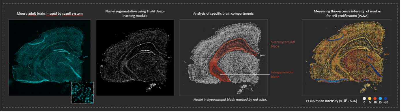
Image analysis of individual cells in an adult mouse brain using the TruAI deep-learning module. The brain sample was prepared by Dr. Jana Krejci (Department of Cell Biology Epigenetics at the Institute of Biophysics).
I used to spend hours at the microscope manually acquiring images, and afterward even more hours at the computer analyzing them. These experiments would take me months to complete. Now, with the scanR, everything can be done in just a few hours.
Evident: How do these applications benefit from the high-content analysis capabilities and image-based cytometry approach of the system?
Hana: In the field of cell biology, I believe that combining unbiased quantitative information with images is a reliable and precise way to represent vital cellular processes. The scanR system offers an elegant and smart solution for this. As I mentioned before, the scanR system provides fully automated acquisition and analysis of samples, enabling us to obtain unbiased and quantitative information about the cellular processes. Importantly, with just one click, one can directly inspect the images that underlie the data.
Additionally, it is possible to create unbiased image galleries that illustrate the dynamics of proteins of interest throughout the life of the cell. Thanks to this unique image-data coupling, it is possible to further fine-tune the analysis of cellular processes to specific compartments, such as the nucleus or cytoplasm, as well as subnuclear compartments such as DNA repair foci, etc. This visual complementation of quantitative data helps to solve biological problems more effectively.
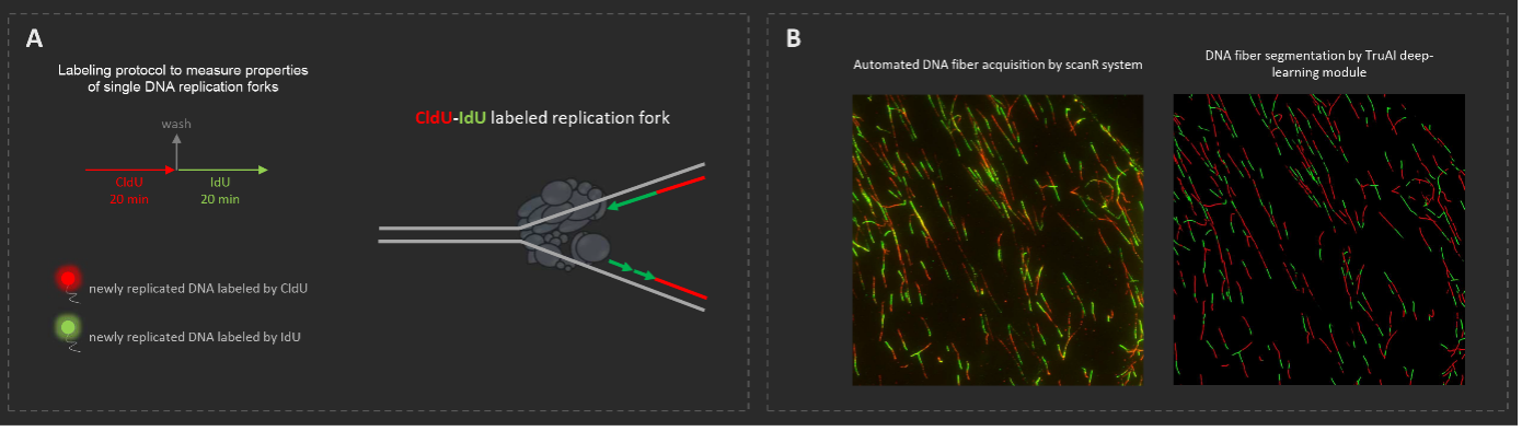
Example of ongoing work on automated acquisition and analysis of DNA fibers by combining the scanR and cellSens™ systems. (A) A CldU-IdU labeling protocol to visualize single replication forks. (B) Examples of automated DNA fiber acquisition and segmentation by the scanR system and cellSens™ software.
Evident: Has the system changed how you approach microscopy as a tool in cell biology and molecular biology? Do you use the TruAI™ deep-learning capabilities of the system?
Hana: During my PhD studies in Professor Jiri Lukas’s lab, I was introduced to high-content imaging and the scanR system. This revolutionary technology completely changed my perspective on cell biology and research in general. I was particularly impressed by its ability to provide an unbiased and fully automated analysis of cellular processes. Given the current reproducibility crisis in scientific research, I firmly believe that researchers should pursue tools that enable them to obtain and analyze data in a fully automated manner, presenting an unbiased representation of cellular processes.
In the current era of artificial intelligence (AI), image analysis has advanced beyond its previous limitations. The scanR system now offers the TruAI deep-learning module, which has expanded the scope of my research. Previously, I focused primarily on analyzing single cells in large populations of cultured human cell lines. However, with the AI module, we can, for instance, conduct extremely precise image analysis of individual cells in organ sections.
Another example of how we can push the limits of our image analysis capabilities is by combining the scanR and cellSens™ systems. This enables us to perform fully automated acquisition and analysis of single molecules, such as DNA replication forks prepared by the DNA fiber technique. These are some examples of new applications we are currently working on in my lab, and we hope that other researchers will benefit from these tools soon to make great discoveries in the field of cell biology.
Evident: What do you like most about the scanR system? How is the reception among your research associates?
Hana: I appreciate many features of scanR system, but if I should highlight just one, then it would be the unique connection between automated acquisition and analysis, enabling us to perform multiparameter analysis of acquired images in real time. I was impressed by this feature when I was a PhD student, and every new user in our small scanR user community at our institute is equally impressed.
One of my colleagues summed it up best when he said, “I used to spend hours at the microscope manually acquiring images, and afterward even more hours at the computer analyzing them. These experiments would take me months to complete. Now, with the scanR, everything can be done in just a few hours.”
For More Information on the scanR System
We extend our sincere thanks to Dr. Hana Polasek-Sedlackova for sharing her experience with the scanR high-content screening station. For more information on the scanR system and our other life science research solutions, contact Evident directly or your local Evident sales representative.
Photo of Dr. Hana Polasek-Sedlackova by Jana Mensatorova.
References
- Polasek-Sedlackova H, Miller TCR, Krejci J, Rask MB, Lukas J. Solving the MCM paradox by visualizing the scaffold of CMG helicase at active replisomes. Nat Commun. 2022 Oct 14;13(1):6090. doi: 10.1038/s41467-022-33887-5. PMID: 36241664; PMCID: PMC9568601. https://www.nature.com/articles/s41467-022-33887-5
- Sedlackova, H., Rask, M.-B., Gupta, R., Choudhary, C., Somyajit, K., Lukas, J. (2020, October 21). Equilibrium between nascent and parental MCM proteins protects replicating genomes. Nature News. https://www.nature.com/articles/s41586-020-2842-3
Related Content
Instance Segmentation of Cells and Nuclei Made Simple Using Deep Learning
Yeast Protein Localization Classified Using TruAI™ Deep-Learning Technology