To keep up with modern research needs, imaging core facilities around the world are looking to increase automation and device integration. With adaptable and advanced tools, researchers can capture a wider range of dynamic cellular processes.
To provide more advanced imaging systems for modern research, equipment suppliers are working closely with customers and other suppliers to develop novel solutions. It is in this spirit of collaboration that we helped create an adaptable imaging and flow control system.*
Dr. Kai Schleicher, advanced microscopy specialist at the Imaging Core facility of the Biozentrum at the University of Basel, Dr. Sebastien Peter, sales specialist at Olympus, and Elveflow, a microfluidic instruments specialist, came together to create a solution that enables coordination of confocal imaging and microfluidic flow control using Olympus’ cellSens imaging software.
In addition to imaging cells in perfusion conditions, this system supports rapid, precise liquid handling—a major benefit for experiments requiring drug treatments or buffer changes.
The Initial Motivation for the Adaptable Imaging and Flow Control System
The project idea arose through a specific request, and Kai immediately saw the many benefits of an adaptable solution that links confocal imaging with microfluidics.
He explained, “A researcher wanted to set up a system that enabled microfluidic devices with several inlets and different pressures to be controlled by microscope software for coordinated imaging. As an imaging facility, it’s important to consider how our systems can adapt to handle a broader range of applications. This project offered an ideal opportunity to use device integration.”
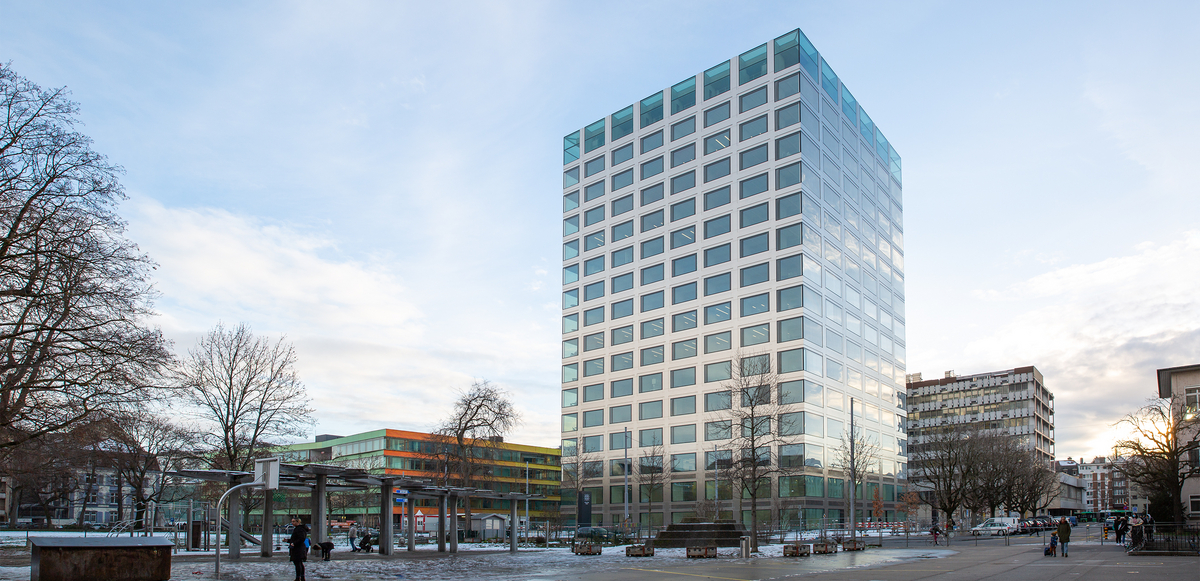
The Biozentrum of the University of Basel
Choosing the Right Equipment to Integrate Microfluidics and Microscopy
To kickstart this project, Kai first contacted Sebastien at Olympus. Kai was already familiar with Olympus microscopes and software. The Biozentrum facility had Olympus IXplore™ microscope systems in place, with the IXplore SpinSR super resolution microscope system proving particularly popular with researchers.
“We decided to collaborate with Olympus because we have a great relationship and they are always helpful,” explained Kai.
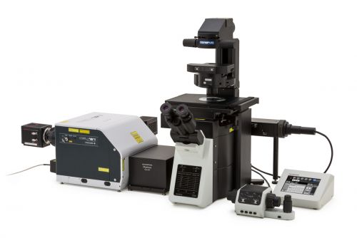
The Olympus IXplore SpinSR super resolution microscope system and real-time controller (RTC)
The flexibility of the IXplore SpinSR microscope system made it ideal for this integrated imaging solution.
Kai explained, “The IXplore SpinSR system has a real-time controller (RTC). This means it can easily interface with third-party devices—making it easy to adapt the system to changing customer needs. The flexibility and ease of use of both the IXplore SpinSR microscope and cellSens software simplify the system usability. The microscope’s high-quality live cell 3D imaging enables a broad range of downstream applications.”
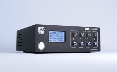
The Elveflow OB1 microfluidic flow controller
The next step was to choose a microfluidic system. Kai and Sebastien quickly chose Elveflow’s OB1 microfluidic flow controller.
“We chose the OB1 microfluidic flow controller as it provides precise control of flow rates on several channels and the flexibility to send and receive signals from the RTC. This enables accurate coordination of imaging and flow control,” explained Kai.
Hardware Setup and Testing for the Imaging and Flow Control System
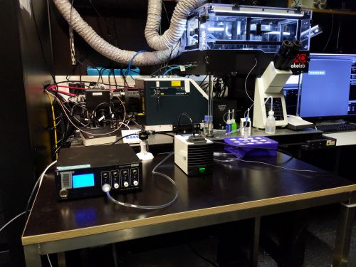
The imaging and flow control system at the Biozentrum’s Imaging Core Facility
With a plan in place, the next step involved hardware setup and testing. Enabling communication between the IXplore SpinSR system and the OB1 microfluidic flow controller was straightforward. It involved connecting the devices with standard BNC cables.
In this setup, sequences written on cellSens software can control the activity of the IXplore SpinSR system and trigger activation of sequences written on Elveflow ESI software—controlling the activity of the microfluidic pump for automated liquid handling.
cellSens software can also receive feedback from external devices such as the OB1 controller. This means users can create simple on/off protocols and more dynamic protocols that involve communication between devices.
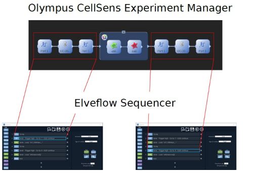
Olympus cellSens software interfaces with the Elveflow sequencer
By writing and running multiple sequences on cellSens software, they were able to quickly confirm that the system was working efficiently.
Kai commented, “The cellSens software is really nice. The simple drag-and-drop interface means that protocols are very easy to write and run. The option to secure general settings and keep them separate from individual user settings is useful to prevent users from accidentally altering sequences that may be commonly used at our imaging facility.”
In only a few hours, they had created an intuitive automated system suitable for a wide range of imaging challenges.
Kai noted, “What’s really great about this combined microscopy and flow control system is its flexibility and how easy it is both to use and set up. The system also requires very little training. This is important as users at our facility want an intuitive system that they can verify is working themselves.”
Enabling Long-Term Research and Scientific Discoveries through Collaboration
Kai believes that collaboration was a key factor in making the integration process so rapid and straightforward.
“The support from Olympus and Elveflow made the process a lot easier. I believe that everyone could achieve the same results as us in the same time frame, without needing strong experience in integrating devices. I would definitely advise anyone to reach out to them for help,” said Kai.
As our understanding of complex biological systems progresses, it’s clear that extending the capability of imaging systems to handle a greater number of advanced applications will be crucial to keep researchers at the cutting-edge of discovery.
Through collaboration with equipment suppliers such as Olympus and Elveflow, imaging core facilities can exploit the synergy of combining their imaging systems with external devices—forming powerful tools that enable novel scientific breakthroughs and help users adapt to the rapidly changing demands of modern research.
Acknowledgements: All Elveflow equipment mentioned in this blog post was kindly provided by Simon Van Vliet and Hector Arturo Gonzalez from the Jenal lab at the Biozentrum of the University of Basel.
Article also published on ElveFlow.com
Related Content
Biofilms in Space: Confocal Laser Scanning Microscopy Helps NASA Research Experiment Take Off
Label-Free Quantification You Can Count On: A Deep Learning Experiment