As our jurors reviewed the hundreds of submissions for our 2019 Image of the Year (IOTY) Life Science Light Microscopy Award, one immediately caught their eye: a vibrant image of a mouse brain slice. Aptly named “Neurogarden” for its garden-like architecture of neurons, this stunning capture won our top global prize.
We chatted with the microscopist behind the image: Ainara Pintor from Spain. She tells us more about her scientific background, the prize-winning image, and why the art of science matters.
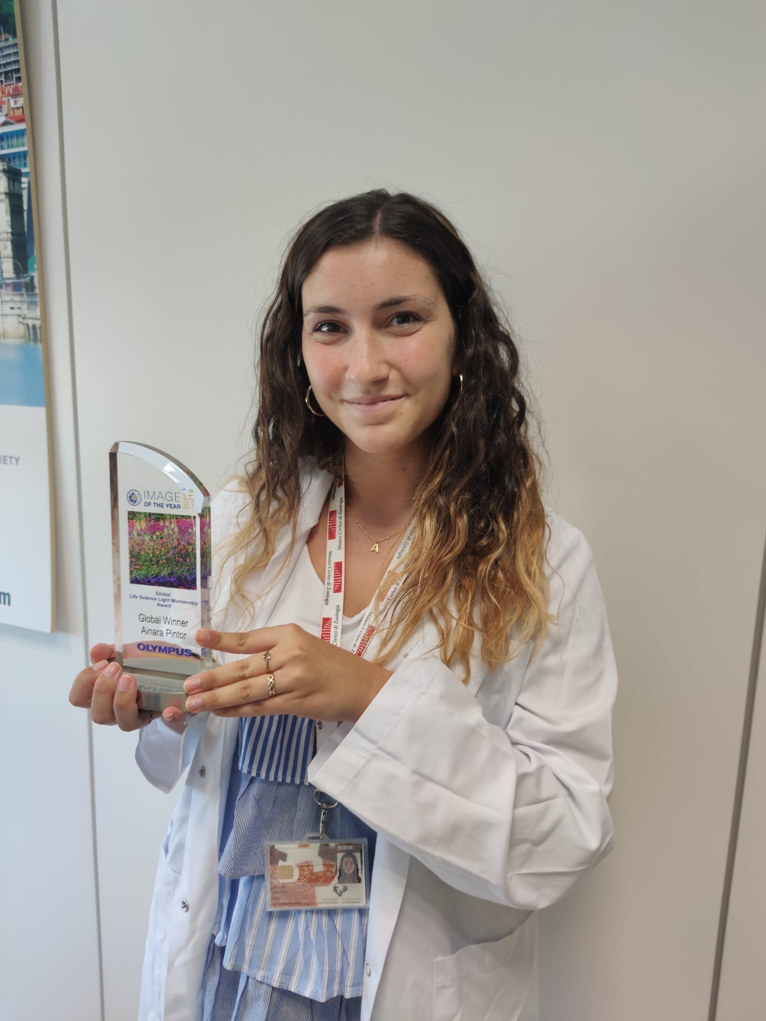
Ainara Pintor shows off her 2019 Global IOTY Award
Q: Can you tell us about your scientific background?
A: I studied Molecular Biology at the University of the Basque Country (Universidad de País Vasco) but concluded my final year at the University of Florence in Italy, where I specialized in neuroscience for the first semester.
Neuroscience was something that I’d always been interested in, and I knew that I wanted to dedicate myself to it. During the second semester, I specialized in medical biology, studying everything from immunology to oncology. After that, I concluded a master’s at the University of the Basque Country in biomedical research, which lasted one year.
Later I joined the Molecular Cognition Laboratory—Shira Knafo's Lab, where I currently work. I started working there in a traineeship and then I was able to get the contract to do my dissertation as a pre-doctoral researcher, which I started in March. I shot this picture during that traineeship when I was finetuning my techniques.
Q: What does your winning image show?
A: This stunning fluorescence image shows the immunostaining of a Thy1-EGFP mouse brain slice with two fluorophores.
In green are the excitatory hippocampal neurons, which express green fluorescent protein under Thy1 promoter. In red are the fat mass and obesity-associated (FTO) protein revealed with Alexa Fluor 594 antibodies. In blue are the cell nuclei labelled with DAPI.
I captured these details with a super-resolution confocal microscope system: the IXplore SpinSR micro spinning disk confocal microscope.
Q: Why did you choose to submit this image?
A: I chose this image because, to me, it seemed that it summarized the exact place where this protein is located within the neuron, which is ultimately our goal.
Also, it’s a special image since it’s not only scientific but also quite inspiring, beautiful, and peculiar. I mean, as a scientist, I know they’re neurons. But if you ask someone who’s not familiar with the field, they might think they're plants facing up or butterflies flying.
In fact, whenever I showed this image to someone outside the laboratory, they would say, “That's nice. It looks like a garden.” So we started to informally call it Neurogarden. And so, the image got its name.
Q: What does the art of science mean to you?
A: To me, it’s important to reflect science as art, because it creates an interest in people who have no scientific background. When someone who has no scientific background asks me “What do you do?” or “What do you do at work?”, it’s difficult for me to explain what I study or what it’s for.
So, by showing them an appealing image like this one, it draws their attention and gives me the opportunity to explain what the image is about and how I got it. It’s a way of creating interest in people who are not dedicated to science.
Q: Where and when did you first learn to use a microscope?
A: I’ve had an interest in microscopy since high school. The first time I used a microscope was during my first year at university when we were observing cell biology. I remember being given samples of tissues that, at first glance, looked like colourful blurs. Ever since then I wanted to know what was really there.
That's when I discovered that I really liked microscopy, because I could see a tiny thing that looked like it had nothing to it—and then look through a microscope and realize it had a whole world inside. The fact that a microscope allows you to see things that the naked eye can’t see fascinated me.
Exploring an image, playing with zooms, and discovering new things through a microscope turned me into a biologist. It's an interest that grew over time.
Q: What do you like about using Olympus microscopes?
A: I’ve used standard optical microscopes, but the Olympus high-resolution one is impressive. My winning image has a very wide depth of field, and I’m dazzled by the image quality. And even more by the speed at which I shot the picture in such a wide depth of field.
It’s important to point out that Olympus cares about reducing phototoxicity. The rotating disk of the microscope I used to shoot this picture reduced phototoxicity and bleaching. The Olympus real-time controller allows you to enhance the device’s speed and accuracy during an automated data acquisition, while the system maintains every frame focused and reduces sample stress during data acquisition.
Q: Is there anyone you’d like to thank for their support?
A: I want to thank my dissertation advisor at Shira Knafo’s. Without her this wouldn’t be possible. She allows me to use her laboratory, teaches me her techniques, and gave me the opportunity to be a member of the group. And I’d also like to thank the Institute of Biophysics and the people who work there.
Get Inspired to Capture Your Own Prize-Winning Image
If you like this image, don’t forget to download a wallpaper version for your desktop or mobile phone! You can do so by visiting our Image of the Year webpage. We hope it encourages you to capture your own art under the microscope.
If inspiration strikes, be sure to submit your images (up to 3!) by January 10, 2021 to our 2020 IOTY contest. Good luck!
Related Content
Artistic Eye View of the Natural World—Meet the 2019 IOTY EMEA Regional Winner
Combining Passions for Science and Art—Meet the 2019 IOTY Americas Regional Winner
Revealing the Beauty of the Microscopic Scale—IOTY’s 2019 AsiaPacific Regional Winner