Lupus nephritis (LN) is a chronic inflammatory kidney disease that occurs frequently (40–60%) in people who have systemic lupus erythematosus (SLE).1 Up to 30% of LN patients develop kidney failure, after which only dialysis or kidney transplant treatment is possible. A goal of lupus research is to accelerate the process of identifying and evaluating LN in biopsied samples to increase the potential for early detection and help improve the disease prognosis.
Challenges of Evaluating Lupus Nephritis (LN) Samples
LN causes a wide spectrum of lesions, which indicates tissue damage. They can be seen by observing kidney biopsy samples under a light microscope. The assessment of kidney lesions and an appropriate classification of LN is outlined in the International Society of Nephrology (ISN)/Renal Pathological Society (RPS) classification. Pathologists use this classification system to determine the stage of the disease.
For example, Figure 1 shows kidney samples of LN class 1 early-stage patients, with no significant glomerular changes compared with healthy subjects. 2
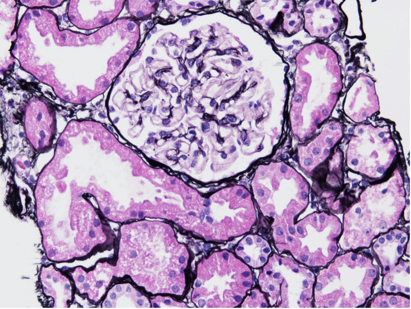
Figure 1. LN class 1 kidney sample biopsy sample, stained with Jonas silver stain.
Whereas in Figure 2, samples from LN class 5 advanced-stage patients show diffuse holes (blue arrow) and small spikes (red arrow) along glomerular basement membranes.3
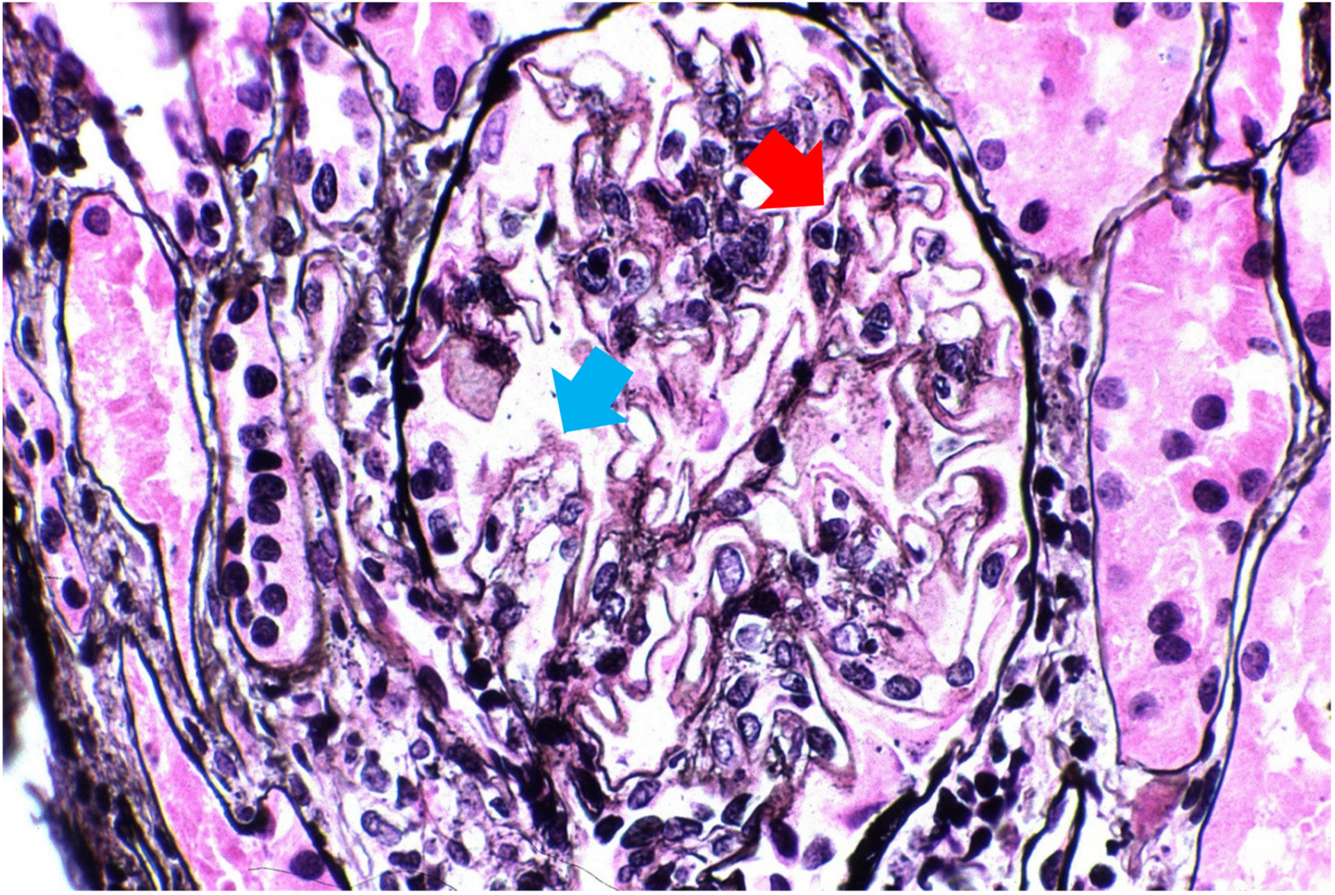
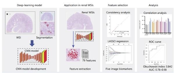
Figure 3. Workflow of MRPS model to perform pathological evaluation using deep learning.
With the help of the SLIDEVIEW™ VS200 research slide scanner, Dr. Shen and a team of researchers acquired 199 high-resolution WSIs of periodic acid-Schiff-stained kidney tissue taken from healthy and LN mice. The images were separated into five different categories by the researchers, with score of 0 given to healthy mice and a score of 4 to mice with severe LN. These images were split into a training set and validation set.
By manually labeling glomeruli and glomerular cells respectively, the researchers were able to use TruAI™ deep-learning technology to generate two neural networks (NNs) from the training set: a glomerular NN and a cellular NN.
The neural networks were then applied to the validation set to extract multiple glomerular and cellular features from each image, as shown in Figure 4.4
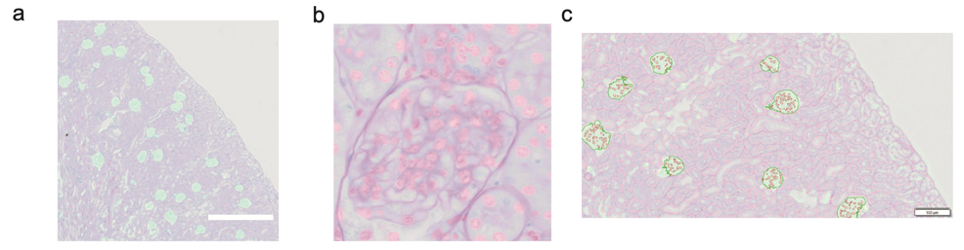
Figure 4. Applying the trained neural networks to a representative validation image. a. All glomeruli were identified in cyan b. All renal nuclei were identified in magenta. c. Identified glomerulus and nucleus were measured in the region of interest (ROI). The glomerular neural network provided an ROI for counting; the cellular neural network counted all nuclei within the ROI.
The perimeter, shape factor, minimum internal diameter, minimum diameter, and the number of objects in each feature are independent predictors that indicate the severity of LN. Based on the parameters above, the researchers developed a scoring model, which performed well in assisting the evaluation and is positively correlated to the score done by researchers.
TruAI™ Deep Learning’s Potential to Assist Image Analysis
This study shows that high-throughput and reproducible analysis of pathological samples is possible using deep-learning technology. This is valuable for basic research where high-throughput WSI analysis is in demand. Deep learning can also be applied to other models and diseases, and it has the potential to significantly reduce researchers’ workloads and increase quantitative precision during analysis.
Acknowledgement:
We thank Dr. Shen Luping for writing the first version of this article, published on Make it Evident丨TruAI 加速狼疮肾炎病理学诊断 (qq.com).
References:
- (PDF) Deep Learning-Based Model Significantly Improves Diagnostic Performance for Assessing Renal Histopathology in Lupus Glomerulonephritis (researchgate.net)
- AJKD Atlas of Renal Pathology: Minimal Mesangial and Mesangial Proliferative Lupus Nephritis (ISN/RPS Class I and II) - American Journal of Kidney Diseases
- AJKD Atlas of Renal Pathology: Membranous Lupus Nephritis, ISN/RPS Class V - American Journal of Kidney Diseases
- Supplementary Material for: Deep Learning-Based Model Significantly Improves Diagnostic Performance for Assessing Renal Histopathology in Lupus Glomerulonephritis (figshare.com)
Related Content
Rapid Automated Detection and Segmentation of Glomeruli Using Self-Learning AI Technology
Perform Accurate and Efficient Microscopy Image Analysis Using TruAI based on Deep Learning