.jpg?rev=287B)
.jpg?rev=7229)
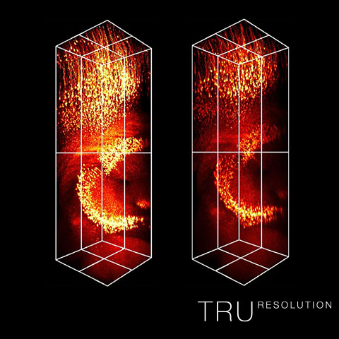
Bright Multicolor FluorescenceIntegrated in FV3000 confocal systems, TruSpectral technology enables much higher light throughput compared to conventional spectral detection units.
| |
|---|---|
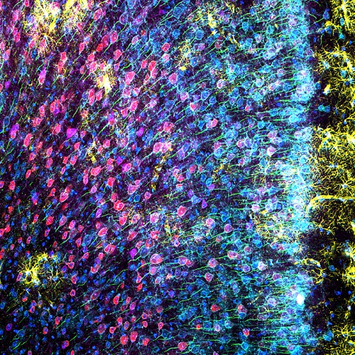
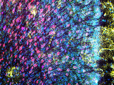
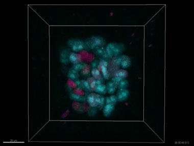
Mouse mPFC labeled with glial fibrillary acidic protein (GFAP; astrocyte marker; yellow), calmodulin-dependent protein kinase II (CaMKII; pyramidal neuron marker; red), amphoterin-induced protein 1 precursor (AMIGO-1; neuronal membrane marker; cyan), parvalbumin (PV; inhibitory neuron marker; purple), ankyrin-G (AnkG; axon initial segment marker; green), and nuclear yellow (nuclei marker; blue). Image data courtesy of Stephanie Shiers, Ph.D. Candidate, Theodore J. Price, Ph.D., Price Lab, Center for Advanced Pain Studies, Department of Neurobiology, School of Behavioral and Brain Sciences, University of Texas at Dallas. |
|
Related Videos | Sensitivity and AccuracyTruSpectral detectors combine high sensitivity with spectral flexibility to detect even the dimmest fluorophores.
|
|---|
Learn More
Multiplexing with the FLUOVIEW FV3000 Confocal Microscope |
3D Time-Lapse Imaging of Spheroids with the FLUOVIEW FV3000 Confocal Microscope |
3D Observation of Cleared Mouse Liver Using the FLUOVIEW FV3000 Microscope |
Development of a New Fucci(CA) Application: A Fluorescent Probe for Visualizing Cell Cycles |
Related Products
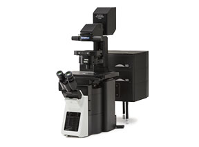
FV3000
|
Innovative Tru technologies help researchers overcome the challenges of imaging experiments.
Seamless deconvolution for clearer, sharper images. |
Stable focus throughout time-lapse experiments. |
Automated correction for brighter and sharper images at depth. |
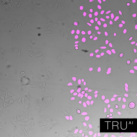
Accurate and efficient image analysis based on deep-learning technology. |
Sorry, this page is not
available in your country.
Sorry, this page is not
available in your country.