Super Resolution and TIRF ImagingOptical techniques such as live cell super resolution and TIRF enable researchers to obtain images beyond the optical diffraction limit. To get the maximum resolution out of an imaging system, it is pivotal that the NA of the objective is as high as possible. |
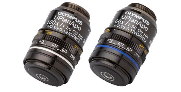
| High-Resolution Objectives for Super Resolution and TIRFGet more out of your samples because we pack more into our objectives. Our proprietary polishing technology has enabled us to create the world’s first plan-corrected apochromat objectives with an NA of 1.5: the UPLAPO60XOHR and UPLAPO100XOHR A Line objectives. Combining these objectives with the IXplore SpinSR system improves the brightness and resolution of your super resolution images. In TIRF (total internal reflection fluorescence) microscopy, these objectives are particularly useful to see surface microstructures. |
|---|
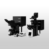
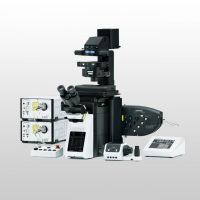
Olympus Super ResolutionThe IXplore SpinSR and FLUOVIEW FV3000 microscopes enable super resolution down to 120 nm using the Olympus Super Resolution (OSR) confocal technique. At the heart of the systems, the high-resolution objective lenses with an NA of up to 1.5 provide the best raw image data to make this possible. Green: Alexa488-labeled Nup358, which localizes to the cytoplasmic surface of a nuclear pore complex |
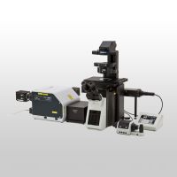
TIRF—Image Dynamic Processes Close to the MembraneAs a pioneer in TIRF microscopy, we offer a diverse line up of A Line objectives with a high NA and magnifications ranging from 60x to 150x. These objectives’ high NA (up to the world’s highest of 1.7) allow unprecedented insights in membrane dynamics. Image data courtesy of Kazuya Tsujita, Ph.D., Toshiki Itoh, Ph.D., Biosignal Research Center, Organization of Advanced Science and Technology, Kobe University Reference: Nat Cell Biol. 2015 Jun; 17 (6): 749–58. doi: 10.1038/ ncb3162. |
 |
Sorry, this page is not
available in your country.
Sorry, this page is not
available in your country.