
Lenses are made of glass, and glass has a different refractive index for each wavelength of light. The lens’ inability to focus different wavelengths of light on one spot is called chromatic aberration. During microscope observation, correcting for chromatic aberration is important for acquiring quality images.
Technical Background
Chromatic aberration is caused by glass that has a different refractive index for each wavelength. Generally, the refractive index of glass is higher for shorter wavelengths. Accordingly, in a convex lens, short wavelength light focuses on a closer position to the lens, and long wavelength light focuses at a distal position from the lens. Concave lenses diffuse shorter wavelength light. Therefore, chromatic correction (CC) is performed by combining convex and concave lenses so that red and blue light focus on the same position below. The unique manufacturing technology of X Line ultra-thin lenses allows more lens elements to be fitted into the objective, leading to unmatched chromatic correction.
Chromatic aberration of single lens |
Chromatic aberration of compound lens |
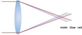
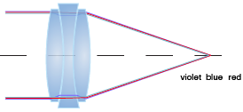
Chromatic aberration comparison of single lens and compound lens.
With lenses manufactured using conventional technology, the chromatic correction of Apochromat objectives ranges from 435–656 nm. Because of this range, the focus position is not colocalized for short wavelengths (< 435 nm) and long wavelengths (> 656 nm). By contrast, X Line objectives have a chromatic correction range of 400–1000 nm while maintaining very high NAs of up to 1.45.
X Line Axial Chromatic Aberration
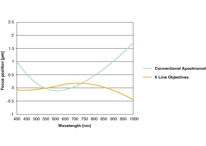
Comparing the focus position in the 400–1,000 nm wavelength range (smaller is better)
Conventional Apochromat Objective |
X Line Objective |
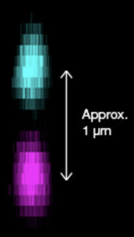
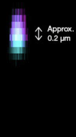
Axial chromatic aberration compared for PSF fluorescent beads (405 nm, 633 nm)
Axial chromatic aberration compared for PSF fluorescent beads (405 nm, 633 nm). Only X Line shows the correct colocalization of the signal.
Your chromatic correction benefits for research imaging:
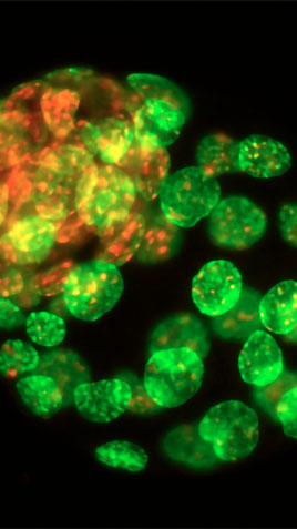
Accurate multicolor images, including near infrared (chromatic correction ranges from 400–1000 nm)
Quantitative and reliable results during colocalization analysis
Enhanced multiplexing capabilities
Your chromatic correction benefits for clinical imaging:
See the true, accurate colors of your sample
Suppress violet aberration to make whites clear and neutral
Improved pink tone for more vivid images with improved contrast
Previous | Next |
 |
Read
|
|---|
Imaging applications
Previous | |||||||||
 Live Cell Imaging | 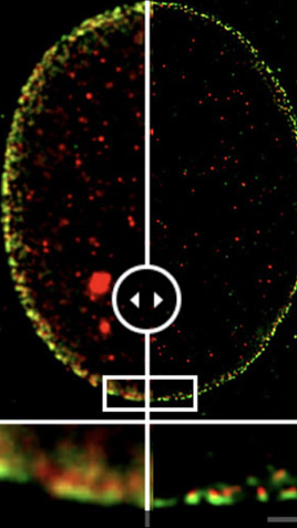 Super Resolution and TIRF Imaging |  Neuroscience and Multiphoton Imaging | 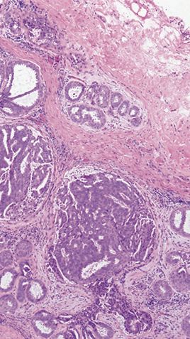 Pathology and Laboratory | 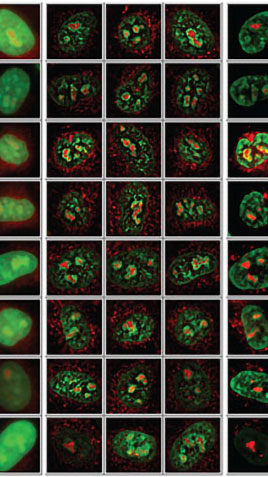 Drug Discovery | 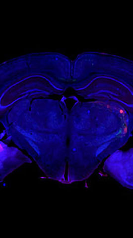 Whole Slide Imaging | 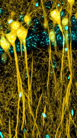 Laser Scanning Confocal |  Cancer Research and Multiplexing |  Fluorescence Imaging |  Organoid and 3D Imaging |
Next |
 |
Sorry, this page is not
available in your country.
Sorry, this page is not
available in your country.