体验4K显微镜的清晰度
4K数字技术引发了显微镜领域的巨大变革,屏幕上图像的清晰度可与传统的显微镜观察相媲美,因此我们能够在屏幕上进行以往需要使用显微镜才能完成的工作。4K显微镜数码相机(DP75、DP28和SC180)所采用的先进技术可呈现前所未有的细节,增强了显微镜用户在屏幕上操作的体验。这项创新技术为用户带来了快速、高效和用户友好的体验,提高了样品评估水平,促进了协同合作,使演示过程生动有趣。
一目了然
· 我们的4K显微镜数码相机配备了强大的CMOS传感器,可进行细节评估,从而提升了在屏幕上工作的效率。
· 栩栩如生的图像吸引了观众,鼓励了协作分析。
· 快速对焦和有效的噪声消除功能,增强了用户在屏幕上完成显微镜工作的信心。
全新的工作方式
多年来,显微镜技术不断地进步和发展。事实上,我们正在见证一个特别有趣的时代:数字技术正在提升成像能力,并为显微镜用户带来诸多好处。
自从数码显微镜相机问世以来,在屏幕上操作就成了一种与通过目镜观察并行使用的有价值的工作方式。然而,在屏幕上操作的要求很高,而且准确的样品分析还取决于相机和图像的多个特性:
- 分辨率:更高的分辨率可捕捉到更高层次的细节,可分析生物样品非常精细的结构。
- 色彩平衡:高保真色彩的呈现对于准确、自信地解读染色模式至关重要。
- 视场:更大的视场有助于在更大的场景中进行有代表性的分析,使用户获得更深入的见解。
- 噪声:低噪声图像即使在光照不足的条件下也能让观察者清楚地看到样品。
- 实时图像速度:无论照明条件如何,都能快速成像,有助于实现快速高效的工作流程,而屏幕上的实时图像在平移过程中也能舒适观察。
为满足这些需求而设计的现代相机得到了不断的改进,最近推出的4K数字成像技术更是实现了图像质量的巨大飞跃。在消费电视领域很常见的4K UHD(超高清)系统包括显示器、相机和软件,具有高分辨率,可以提供比以往更大、更细腻的图像。除了提高的分辨率,4K相机还采用了其他先进的技术,获得了前所未有的图像质量。4K数字成像为显微镜领域带来了大量激动人心的机遇,增强了样品分析和展示效果。
在屏幕上分析的价值
屏幕可视化和通过目镜观察,各有优点和局限性。虽然目镜无疑可以提供逼真的样品图像,但长时间工作会让人感到缓慢和不舒服,而屏幕可视化则提供了一种互补且舒适的替代方案。事实上,4K显微镜现在已经将图像质量提升到一定标准:在数字图像中捕获的信息与通过目镜看到的信息不相上下。
用户越来越多地使用这两种可视化模式,减少了用目镜确认观察结果的需要,从而节省了时间。
虽然“4K”一词描述的是图像分辨率,但通过其他数字技术也能增强图像的质量。其中包括色彩再现,这种技术已发展到可以生成逼真的色彩轮廓以进行准确的分析,特别是在评估组织学样品染色的细微色调时。降噪技术的改善也使得隐藏的细节变得清晰可见。例如,Evident的智能图像平均(OSIA)功能可完全消除图像噪声,既不会降低帧速率,也不会产生伪影,而且无需对传感器进行主动冷却(图1)。这种相机可在各种成像条件下捕捉更多细节,从而适用于许多传统上属于冷却相机领域的应用。因此,科学家们现在可以依靠单个相机来替代专门的低噪声相机,从而提高了成本效益。
除了提高图像质量,数字功能还增加了相机的实用性,考虑到了快速导航到感兴趣的区域并轻松对焦的需要。每天评估大量样品时,速度和舒适度尤为重要,而快速实时图像可在平移过程中保持流畅的移动,非常适合跨样品之间的导航。此外,由于经常需要对组织样品的不同层次进行对焦,因此在分析许多区域时,不断调整焦距以找到所需的设置会非常耗时。使用一种称为“对焦峰值”的新技术可以节省时间(图2)。该功能可直接突出显示实时图像中的所有聚焦区域,这样用户就无需为了找到所需的焦点而对同一区域拍摄多张快照。
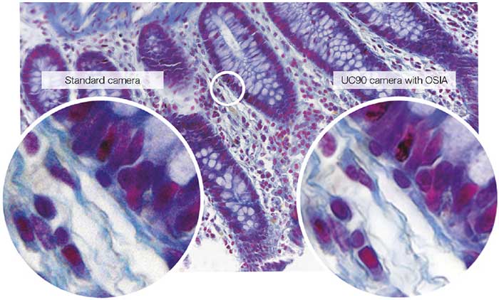
采用Evident智能图像平均(OSIA)技术的主动降噪功能,可以捕捉到原本会因噪声而模糊的细节。
(样品:人类结肠)
科学家可以利用对焦峰值技术快速轻松地选择所需对焦的区域(对焦区域显示为红色)
高效的合作和引人入胜的演示
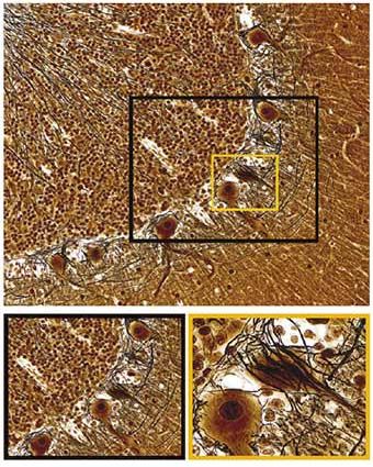
成功的工作流程依赖于高效的合作,而日常工作也需要高效合作来确保决策的准确性。因此,同事之间高效共享详细而准确的信息至关重要。4K显微镜具有更高的分辨率和更大的视场,有助于进行联合图像评估,由于还能在屏幕上显示样品结构的细节和背景,从而增强了演示效果。如图3所示,脑组织切片等大图像提供了具有代表性的样品视图,使复杂的组织结构清晰可见。同时,4K分辨率捕捉的细节还可使图像放大,显示神经元形态的更多细节,以便在组织和细胞层面进行分析和讨论。可以向多位科学家展示大量信息,从而通过屏幕上的图像使整个团队全面了解研究的进展情况。为了分析更大的区域,还可以使用数字图像拼接功能,进一步扩大视场,将来自样品多个区域的多个采集图像合并为一张高质量的明场全景图像。4K显微镜加快了这个过程,使得为生成最终全景图像所需的单次采集次数大大减少。通过这些功能,4K显微镜捕获的栩栩如生的图像还能让科学家们在会议讲座或教学过程中生动地展示科研成果,调动所有观众的兴趣。
在屏幕上显示图像在许多情况下都是有益的,例如大学讲师在研讨会上使用显微镜提供的实时图像时。当演示者在样品上平移并放大某个区域,以在大屏幕上显示微小的细节时,快速成像功能有助于流畅的导航,为观众快速显示感兴趣的区域。与在教学环境中使用多头显微镜相比,一大群科学家只需使用一台配备了4K相机和4K大屏幕的标准显微镜,就可以轻松清晰地观察和讨论样品。
为了更全面地了解样品,大视场有助于在单个图像的背景中分析样品,而高分辨率则可以放大图像以显示详细的结构。
总结
现在,数字成像技术已经发展到可以通过显示器对样品进行准确、可靠分析的水平。有了先进的4K显微镜相机(例如Evident的DP75、DP28和SC180),就可以将改进的分辨率和视场与尖端数字功能相结合,进行深入分析和清晰交流。除了提高屏幕图像质量之外,还可以通过快速实时成像和快速对焦节省时间,同时可以直接在屏幕上确认观察结果,只需偶尔通过显微镜目镜看一眼即可。在4K显微镜时代,图像变得栩栩如生,可使显微镜用户以前所未有的方式探索他们的样品。
适于这类应用的产品
对不起,此内容在您的国家不适用。