Introducing the FV4000 Laser Confocal Microscope
Transforming Precision Imaging
The metamorphosis is complete.
The next evolution of confocal imaging has arrived with the FLUOVIEW™ FV4000 microscope, and this time, we’re transforming how you think about confocal microscopy.
Powered by our pioneering new SilVIR™ detector, you can achieve results that you’ll need to see to believe. Don’t take it from us—see what your colleagues who’ve tried the system are saying.
"Compared to other confocals I have experience with yours was very stress-free to use (no fear of destroying the sample or feeling overwhelmed with the software/general operation) and required a minimal amount of adjustments from the user in order to produce good quality data without feeling that my options are limited.”
| “The detectors are easy to set up due to their high dynamic range. They can handle low signals, but also do not switch off and perform well with high signals. This makes it very user friendly to set up the microscope. The photon counting mode is a highly appreciated function. The high precision, simple adjustment, and high flexibility make the FV4000 a very valuable tool for imaging facilities with users of different experience levels and varying complexity in their imaging experiments.”
|
.jpg?rev=AC90)
.jpg?rev=AC90)
Don’t take their word for it.Experience the FV4000 microscope’s abilities for yourself, with innovations that include:
| ||
The image captured using the SilVIR detector has extremely low background noise compared to the image captured using a GaAsP-PMT.
| Instead of choosing to focus on either dim or bright fluorescence areas, the FV4000 microscope can capture both in one image without saturation or loss of information thanks to the SilVIR detector’s high dynamic range.
|
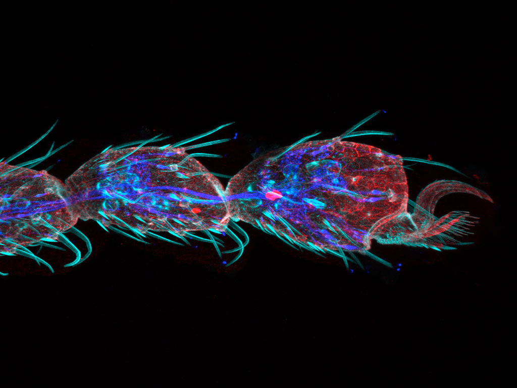
Tip of a Drosophila leg (42-hour pupation), stained with phalloidin (AlexaFluor 405, F-actin, Cyan), anti-phosphotyrosine antibody (AlexaFluor 555, cell surface, red), and anti-HRP antibody (AlexaFluor 647, axon, blue). Sample Courtesy of: Zhengkuan Sun, Shigeo Hayashi, Laboratory for Morphogenetic Signaling, RIKEN Center for Biosystems Dynamics Research, Japan. |
“Together with the AI restoring tools, it gives a perfect combination for fast and high-quality volumetric imaging.”Edwin Hernandez | Core Facility Manager, Cajal International Neuroscience Center (CINC) | “The new FV4000 will allow for faster imaging and better preservation of the sample and will allow better reproducibility.”
|
.jpg?rev=AC90)
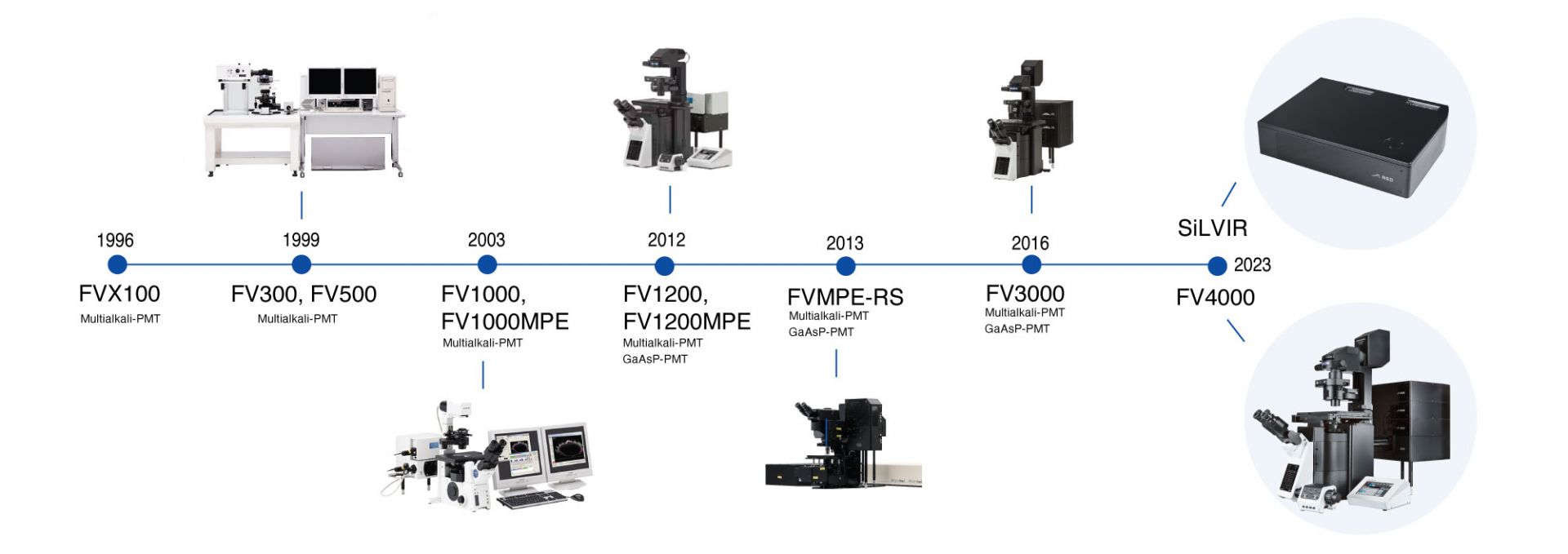
Laser Confocal Microscope Timeline
“Demonstratively better signal to noise [than other systems].”
| “I am really impressed with the system and its performance. The super sensitivity was really impressive.”Sara R. Roig | Advanced Microscopy Specialist, University of Basel |
.jpg?rev=AC90)
Sorry, this page is not
available in your country.
Sorry, this page is not
available in your country.