Confocal Laser Scanning ImagingLaser scanning microscopy (LSM) is used to obtain high-resolution, high-contrast imagery of a sample. LSM can scan samples point by point, resulting in optical sectioning that can be used to construct precise 3D images. |
| High Resolution and Large Field of View from a Single ObjectiveCover a magnification range from 40X to 100X without changing objectives using a 40X, 1.4 NA X Line objective and the FV3000 microscope's optical zoom. |
|---|
Better Stitching in Multicolor Panoramic ImagesImproved image flatness enables you to acquire superior stitched images with less zooming in a wide wavelength range from 400 to 1000 nm. Brain section of Fucci2 transgenic mouse |  |
|---|
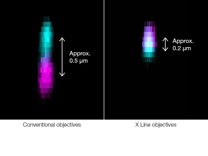 | True Colocalization AnalysisX Line objective’s chromatic aberration correction from 400 to 1000 nm is the basis for accurate quantitative colocalization analysis. Trust what you see—only with X Line objectives can you can be sure that two signals really colocalize, as insufficient chromatic correction can easily introduce a shift of fluorescence channels in the Z dimension. Image: HeLa cell labeled by FISH technique CEP17(Spectrum Green), CEP18(Spectrum Orange), Nuclear (DAPI)* When observing with conventional objectives, signals located at the bottom of the cell’s nucleus appear outside the nucleus. Scale bar: 2 μm |
|---|
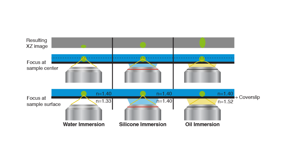
See the True 3D Shape of Your Sample with Silicone Immersion ObjectivesMatching the refractive index of a sample and immersion media is very important to get accurate 3D images. Mismatches of refractive indices (water or standard immersion oil) will lead to distortions of the 3D image. Only silicone immersion oil provides the right refractive index of living samples and results in true 3D images. Olympus’ A Line silicone oil objectives enable you to see the truth in your 3D samples. |
|
|---|
 |
Sorry, this page is not
available in your country.
Sorry, this page is not
available in your country.