
Image flatness is an important indicator of the reliability of quantitative analytical data from observations that require high resolution over a large field of view (FOV). Spherical and coma aberration, astigmatic aberration, the effect of field curvature, and peripheral darkening are all factors that can limit an objective’s image flatness.
Technical Background
With X Line objectives you can now discover unparalleled flatness from the center to the periphery that doesn’t sacrifice NA or chromatic correction—even in large FOV observations.
Conventional PLAPON60XO |
X Line UPLXAPO60XO |
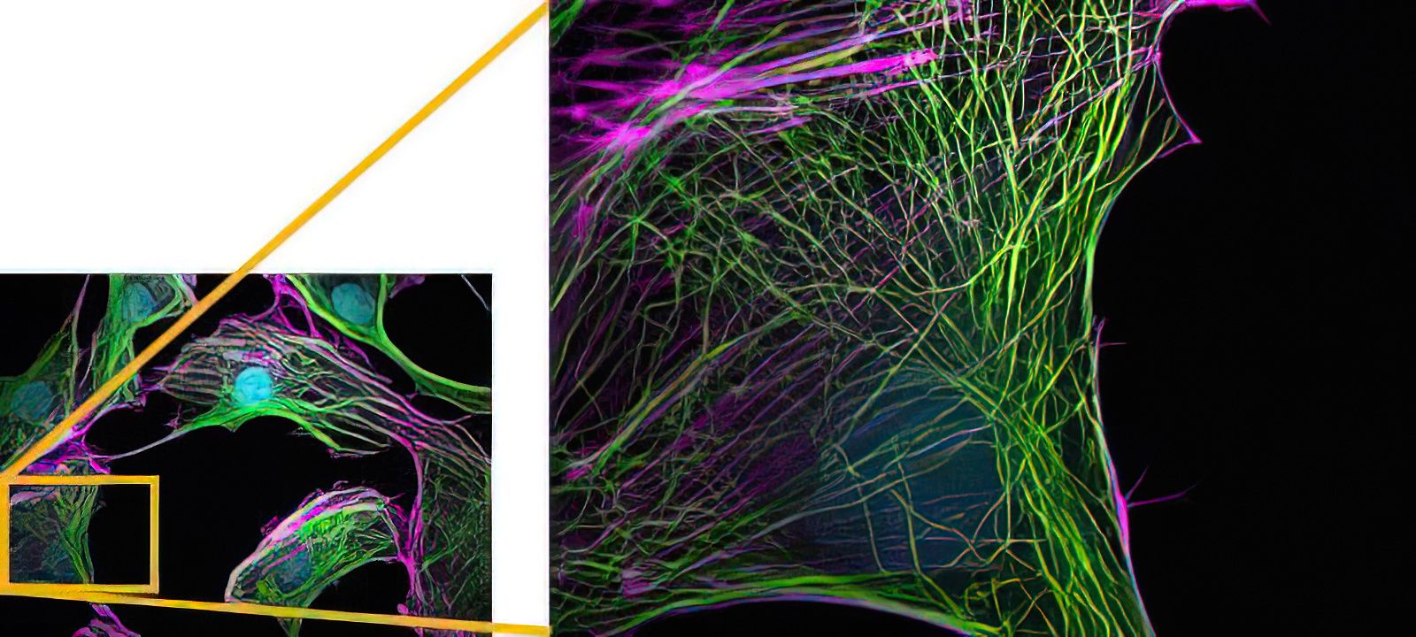
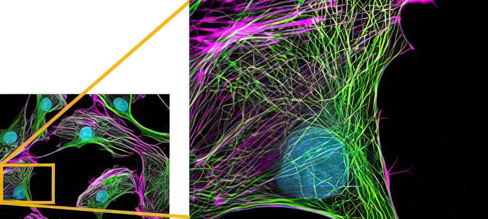
Comparing the image flatness between the conventional PLAPON and the X Line UPLXAPO objectives
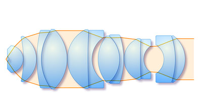
A precise combination of convex and concave lenses is required to reduce field curvature and create flat images.
|
|
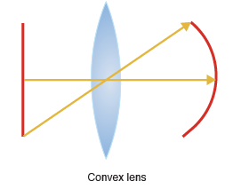
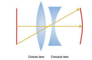
Field curvature through convex and concave lenses
Only a high number of corrective lens elements allow sharp, high-resolution images from the center to the edge of the field.
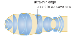
X Line objective’s ultra-thin lens design allows for more corrective lens elements in a limited space, enabling unmatched flatness.
Conventional design (7 groups, 13 lenses) |
X Line ultra-thin lens design (9 groups, 15 lenses) |
Accurate quantitative results over the complete field of view
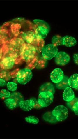
Quantitative analysis and imaging with a large FOV are common thanks to advances in digital technologies. For your analysis to be reliable, high-quality raw data are essential. The images below shows fluorescence of nuclei stained with DAPI (405 nm excitation wavelength) acquired with different objectives and analyzed to automatically recognize nuclei using analysis software. Peripheral nuclei are not accurately recognized in the image because the image flatness is poor. However, using the X Line objective (UPLXAPO60XO; NA 1.42) with excellent flatness, even peripheral nuclei are accurately recognized..
|
|
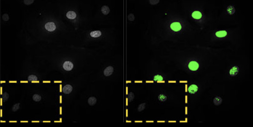
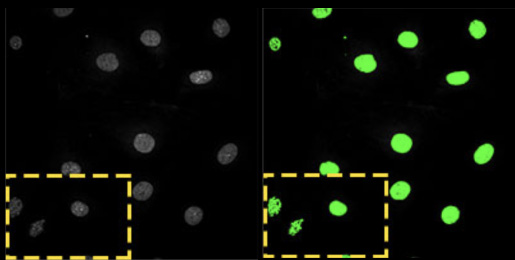
Comparing images at 405 nm (Left: Raw image, right: analysis results)
Your benefits from great image flatness for research imaging:
Uniform image quality from the center all the way to the edge
Correctly detect and analyze objects at the edges
Stitch images together without a shading pattern
Efficient image acquisition with large FOV detection (e.g., sCMOS camera)
Your benefits from great image flatness for clinical imaging:
Edges of your large field of view will stay in focus
Uniform image quality from the center all the way to the edge
Efficient image acquisition for panoramic imaging with less cropping
Next BenefitChromatic Correction |
 |
Read
|
|---|
Imaging applications
Previous | |||||||||
 Live Cell Imaging |  Super Resolution and TIRF Imaging |  Neuroscience and Multiphoton Imaging |  Pathology and Laboratory | 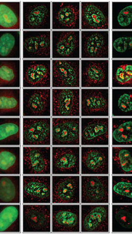 Drug Discovery | 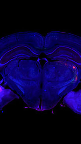 Whole Slide Imaging | 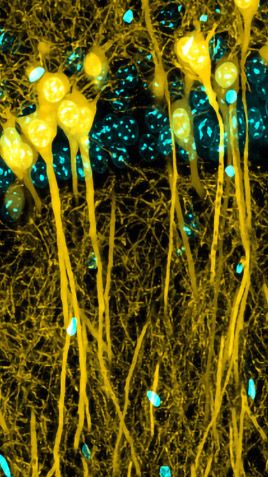 Laser Scanning Confocal | 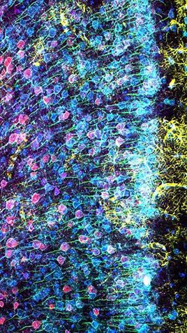 Cancer Research and Multiplexing | 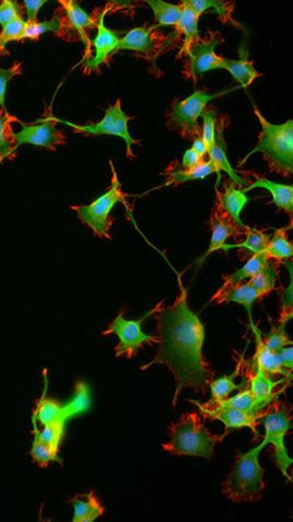 Fluorescence Imaging | 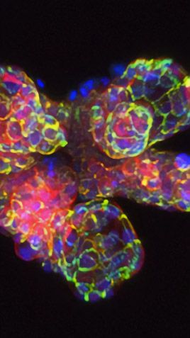 Organoid and 3D Imaging |
Next |
 |
Sorry, this page is not
available in your country.
Sorry, this page is not
available in your country.