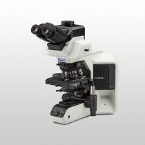
Pathology and LaboratoryIn pathology and laboratory applications, specimens taken from cells, tissues, or organs are examined to study changes in relation to disease. In microscopy, image quality is paramount as it increases efficiency and shortens time to results. This quality depends on the very heart of the microscope—the objective. |
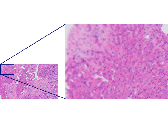 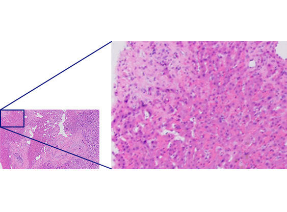 | High Quality from the Center to the EdgePairing our BX53 upright microscope with X Line objectives greatly improves the overall quality of images. See all the details with improved flatness and uniform high quality from the center all the way to the edge. A high numerical aperture (NA) enables X Line objectives to gather more light for brighter, higher-resolution images. |
|---|
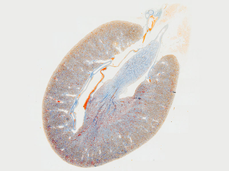
EFFICIENT IMAGE STITCHINGStitching enables you to produce images of whole tissue sections, but if the image flatness is poor, the stitched image can be blurry and present a "patchwork" artifact. X Line to the rescue! These objectives provide uniformly flat images from the center to the edge, enabling you to acquire uniform, high-definition stitched images. The wide field of view allows you to create a large stitched image using fewer images than before, improving efficiency and saving you time. |
|
|---|
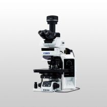
 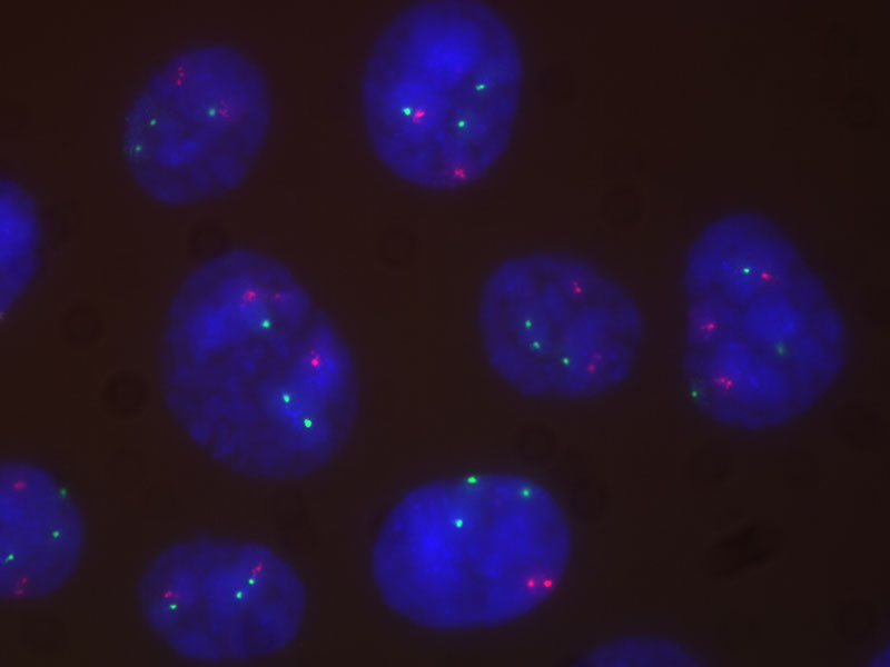 | Improved Images for Clinical ResearchImproved flatness, numerical aperture, and chromatic aberration combine to deliver clear, high-resolution images with excellent color reproduction. The objectives' superior chromatic aberration management delivers better color accuracy across the entire spectrum. The elimination of violet color aberration creates clear whites and vivid pinks, improving contrast and sharpness. Chromatic aberration correction in a wide wavelength range (400–1000 nm) enables you to acquire true, high-resolution, multicolor images during fluorescence observation, such as fluorescence in situ hybridization (FISH). |
|---|
 |
Sorry, this page is not
available in your country.
Sorry, this page is not
available in your country.