
This month’s selection took us from the Amazon rainforests to the canals of Venice, showing the beauty of a range of microscopic subjects across the world.
"Charles Darwin wrote, 'Endless forms most beautiful and most wonderful have been, and are being, evolved.'
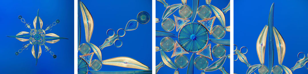
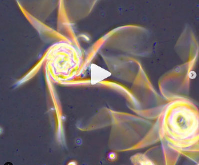
Diatoms are microscopic single-cell algae housed in beautiful glass shells. There are hundreds of thousands of varieties of diatoms all with unique forms. Diatoms range in size from 5 microns to 200 microns (a micron is one-thousandth of a millimeter), so a diatom arrangement of 100 forms would fit inside a punctuation mark of an average-sized text. The image above shows a Victorian-style diatom arrangement made by one of the few living practitioners of this art form, Klaus Kemp. The first diatom arrangements date back to the early 1800s, but the art form reached its peak in the latter part of the century. It was a period of intense interest in the natural world and also a time when the arts and sciences were more closely aligned. Diatom arrangements are a stunning example of that Victorian desire to bring order to the world and to display nature in a rational way. Diatoms can potentially be found in any body of water such as ponds, lakes, rivers, or the sea. After the diatoms have been cleaned, they are placed on a glass slide with adhesive. They can then be manipulated under the microscope until the glue dries. Once the glue has set, a mountant is added that allows the diatoms to be seen more clearly. Imagine the number of hours of work this type of arrangement takes to make!
This photograph was captured using an Olympus X Line 20X objective to achieve as high a resolution as possible. A stitch of 9 different stacks using a total of 416 images was used to produce the final image, which has a resolution of 210 megapixels. I have included a few crops with more details. Diatoms are among my favorite subject to photograph due to their variety and beauty.”
Caption and Image courtesy of Håkan Kvarnström.
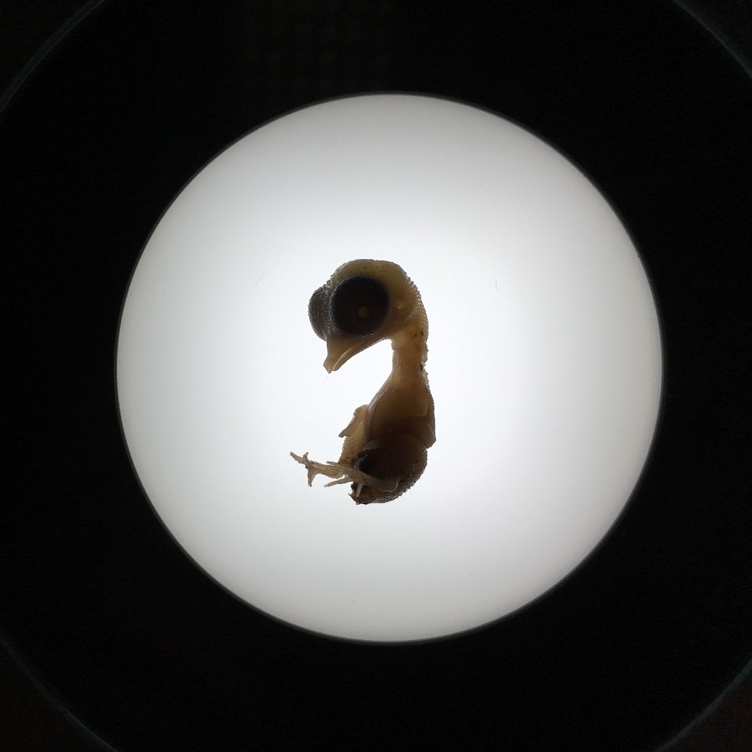
Which came first, the chicken or the egg? In this case, we are looking at both! This image shows a chicken embryo on the ninth embryonic day. It generally takes 21 days of incubation for the embryo to fully develop into a chick and hatch from its shell.
Image captured by Eva Petrovova on an Olympus SZ61 microscope. 2019 Image of the Year Award Submission.
Submissions are now being accepted for our 2020 Global Image of the Year Award Competition. Click here to learn more and see and how you can win a SZX7 stereo microscope with DP27 digital camera or CX23 microscope.
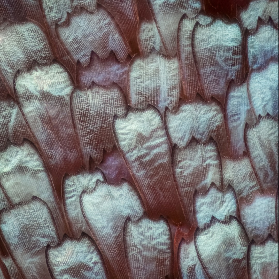
This beautiful image shows the scales of a Panacea prola butterfly. Native to South America, you can find these mesmerizing turquoise blue butterflies frolicking in foliage high in the rainforest in groups of 50 or more. The underside of their wings is bright red, and the vivid colors earn them the common names “prola beauty” and “red flasher.”
Image courtesy of Marco Jongsma. Captured on an Olympus BH2 microscope at 55x magnification.
Everyone loves tardigrades! Here are three facts about water bears that you may not know:
- Tardigrades can withstand environments as cold as -328 °F (-200 °C) or as hot as more than 300 °F (149 °C).
- To survive extreme conditions, a water bear can curl its head and legs into a dehydrated ball called a tun and can be revived if re-introduced to water.
- Tardigrades can produce a protein that protects their DNA from radiation damage.
Image courtesy of Hunter Hines. Captured on an Olympus BX53 microscope with a DP72 camera.
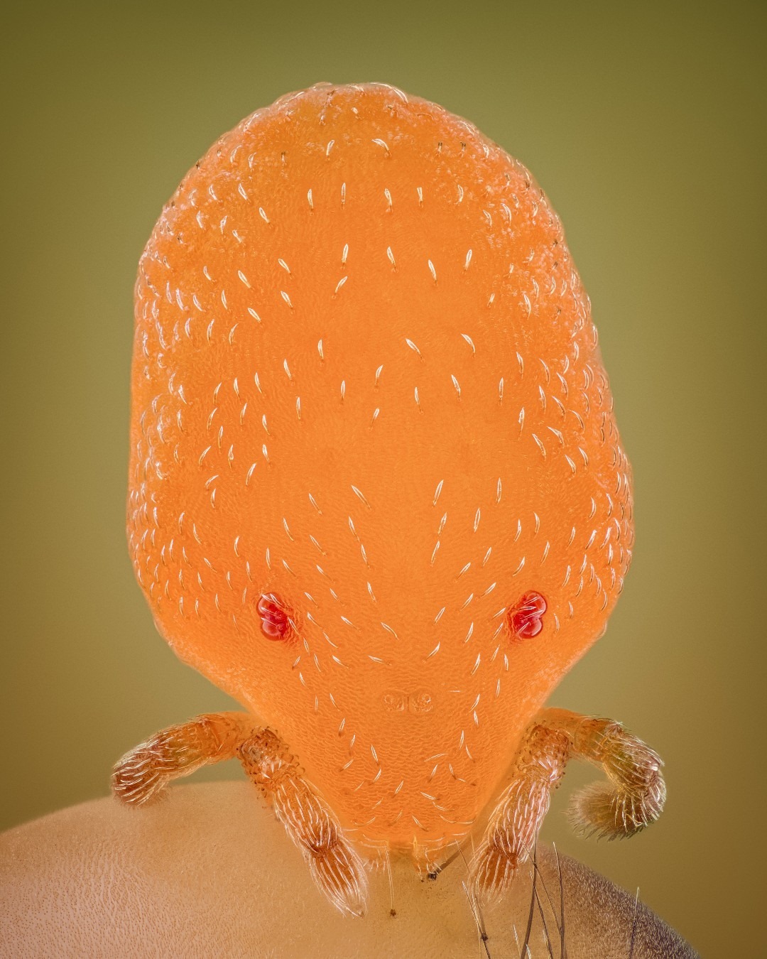
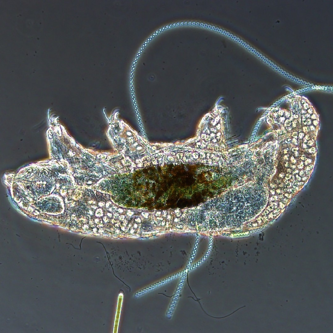
Calyptostoma is a genus of mites in the family Calyptostomatidae with about six known species. If you look closely, you can see that this parasitic mite has attached itself on a cranefly's thorax.
Image courtesy of Leonardo Capradossi. 2019 Image of the Year Award Submission.
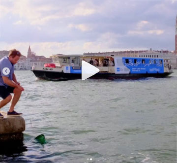
In addition to the above popular images, this past month we featured an Instagram takeover by the talented Benedikt Pleyer.
Benedikt describes himself as “passionate about documenting the microscopic realm all around the world” and took us on a journey through the canals and waterways of Venice, Italy. From diatoms to jellyfish, his takeover showcased both the underwater and images of the city where he collected samples.
Videos courtesy of Benedikt Pleyer. All images captured on an Olympus CX43 microscope.
To see more images like these, be sure to follow us on Instagram at @olympuslifescience!
Interested in sharing your own images?
Visit our image submission site or enter them into our 2019 Global Image of the Year contest.
Related Content
Combining Passions for Science and Art—Meet the 2019 IOTY Americas Regional Winner
See What the Buzz Is About: Our Most Popular Microscope Images for August 2020
