Olympus Image of the Year Award 2020Image of the Year Award 2020 has come to a close. Have a look at below to see the beautiful images we received this year. For the latest information and submission details, click here! |
|
The Global Winner
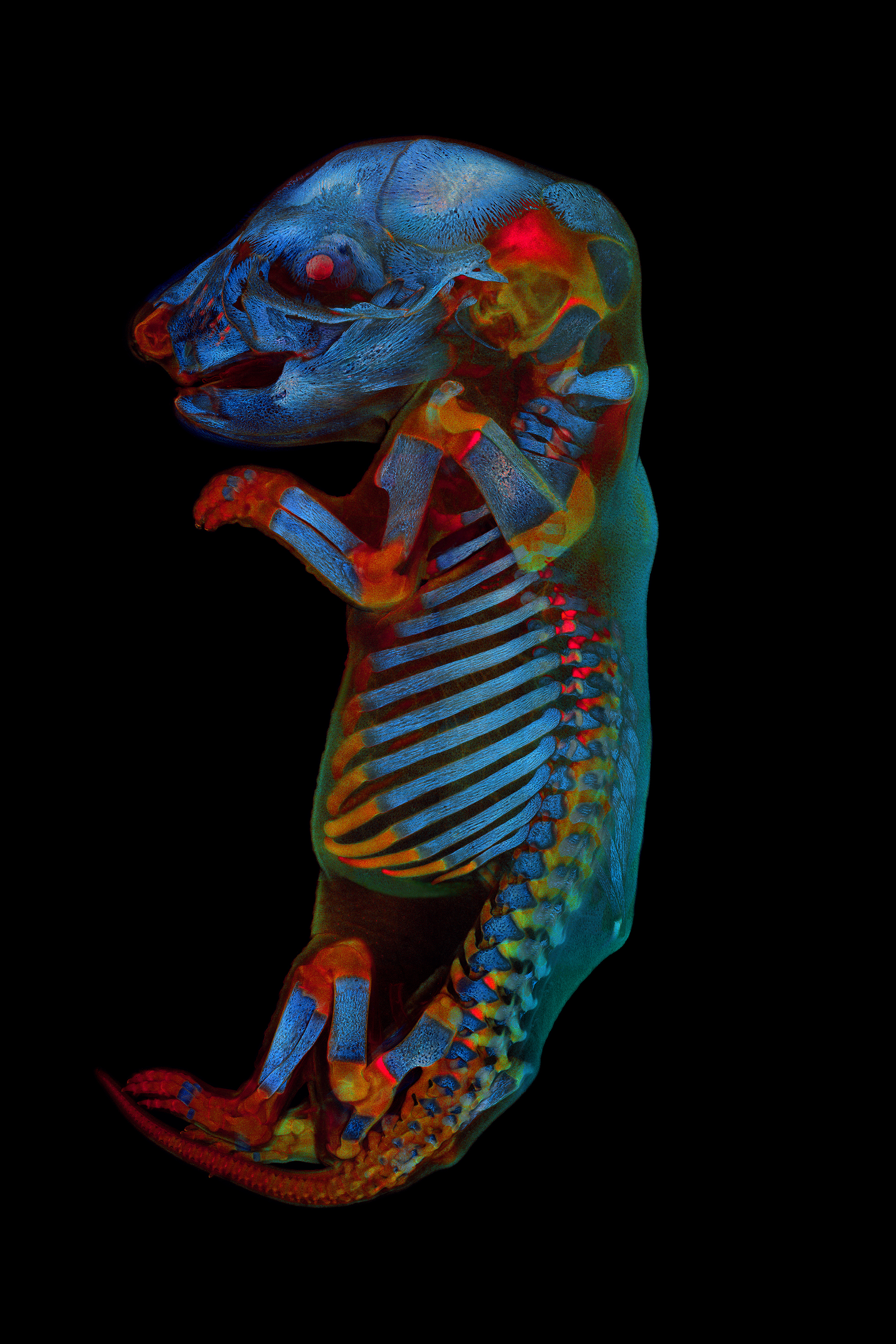 | The winning image was taken by Werner Zuschratter (Germany). Whole rat embryo three channel large scale confocal image of a fixed and cleared rat embryo. Two channels show different autofluorescence sources of the tissue, whereas the third channel shows the skeleton stained by alizarin red. |
Regional Winners
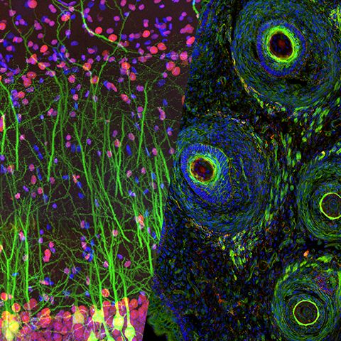
Americas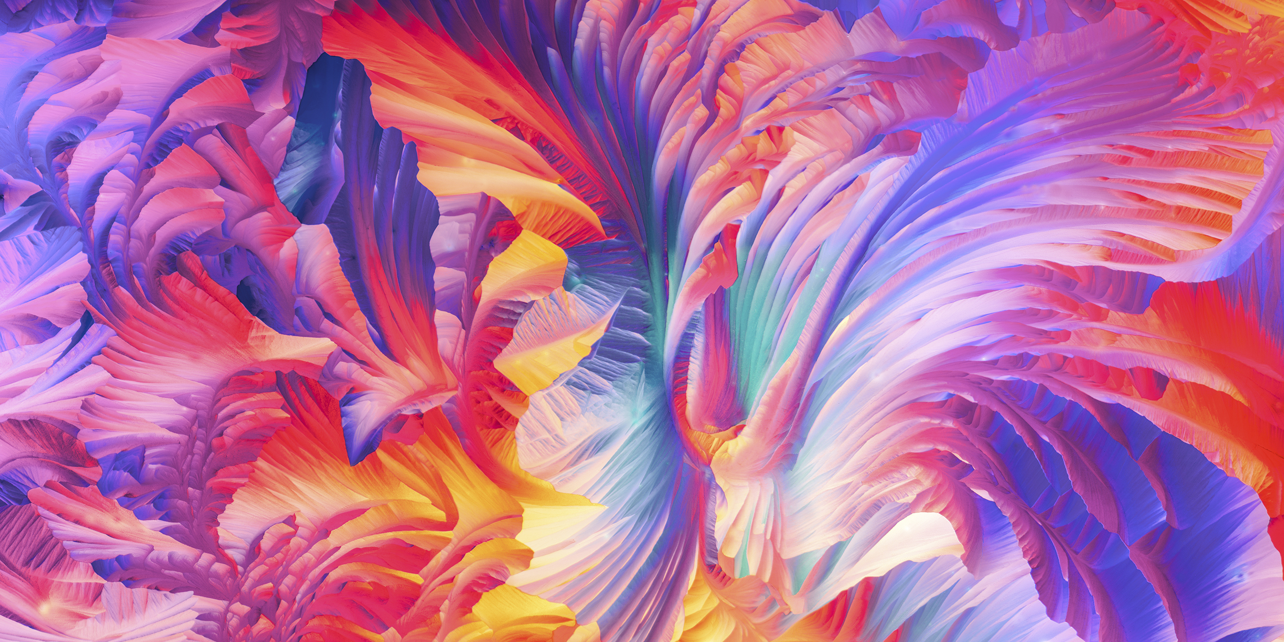 The winning image was taken by Justin Zoll (U.S.A.). Polarized light microscopy panorama of l glutamine and beta alanine crystals. | Europe, the Middle East and Africa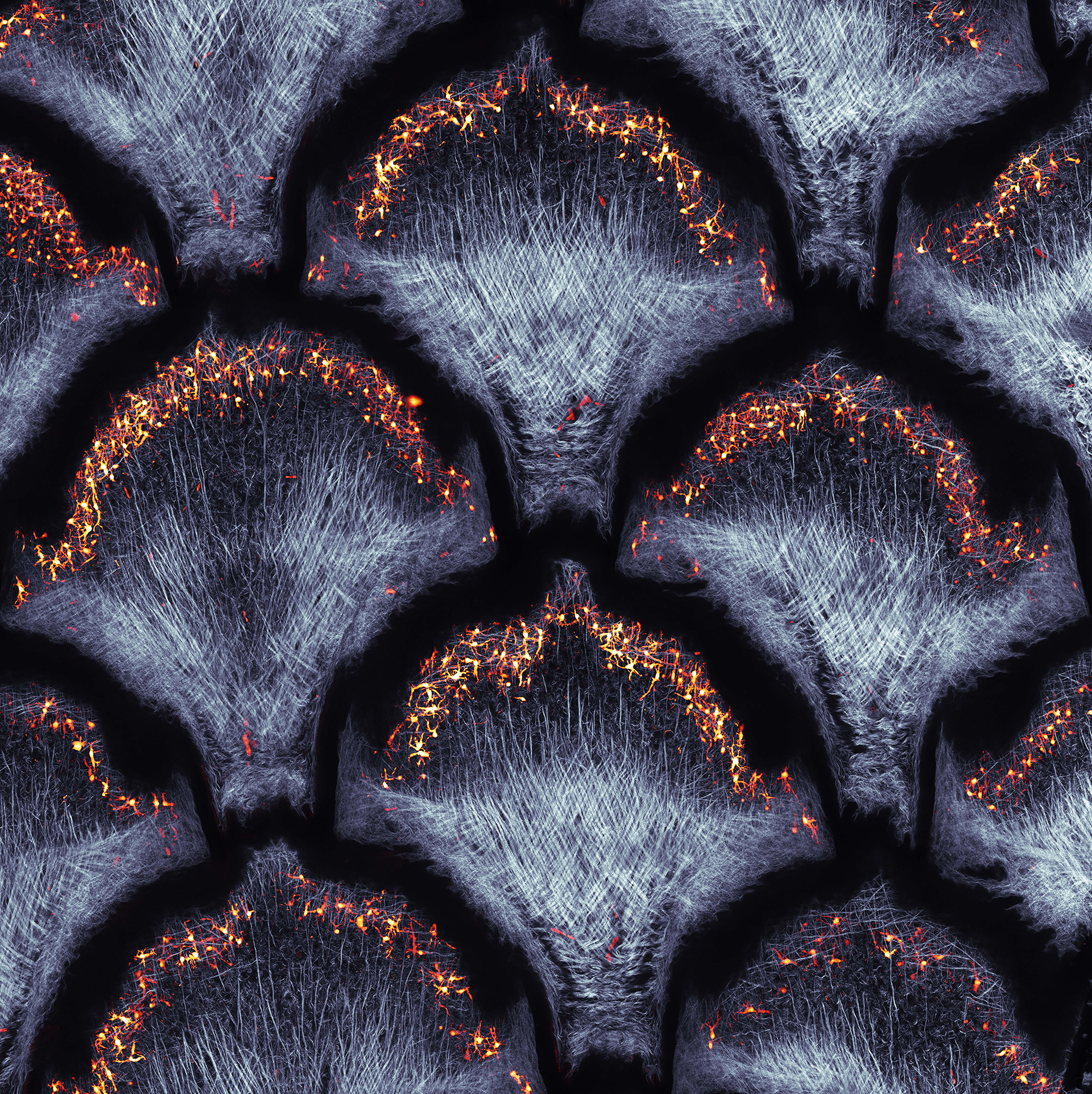 Grigorii Timin (Switzerland)'s image won the EMEA regional prize. Collagen fibers (second harmonic generation) and dermal pigment cells (autofluorescence) in African house snake embryonic skin; maximal intensity projection of 10 confocal slices. | Asia-Pacific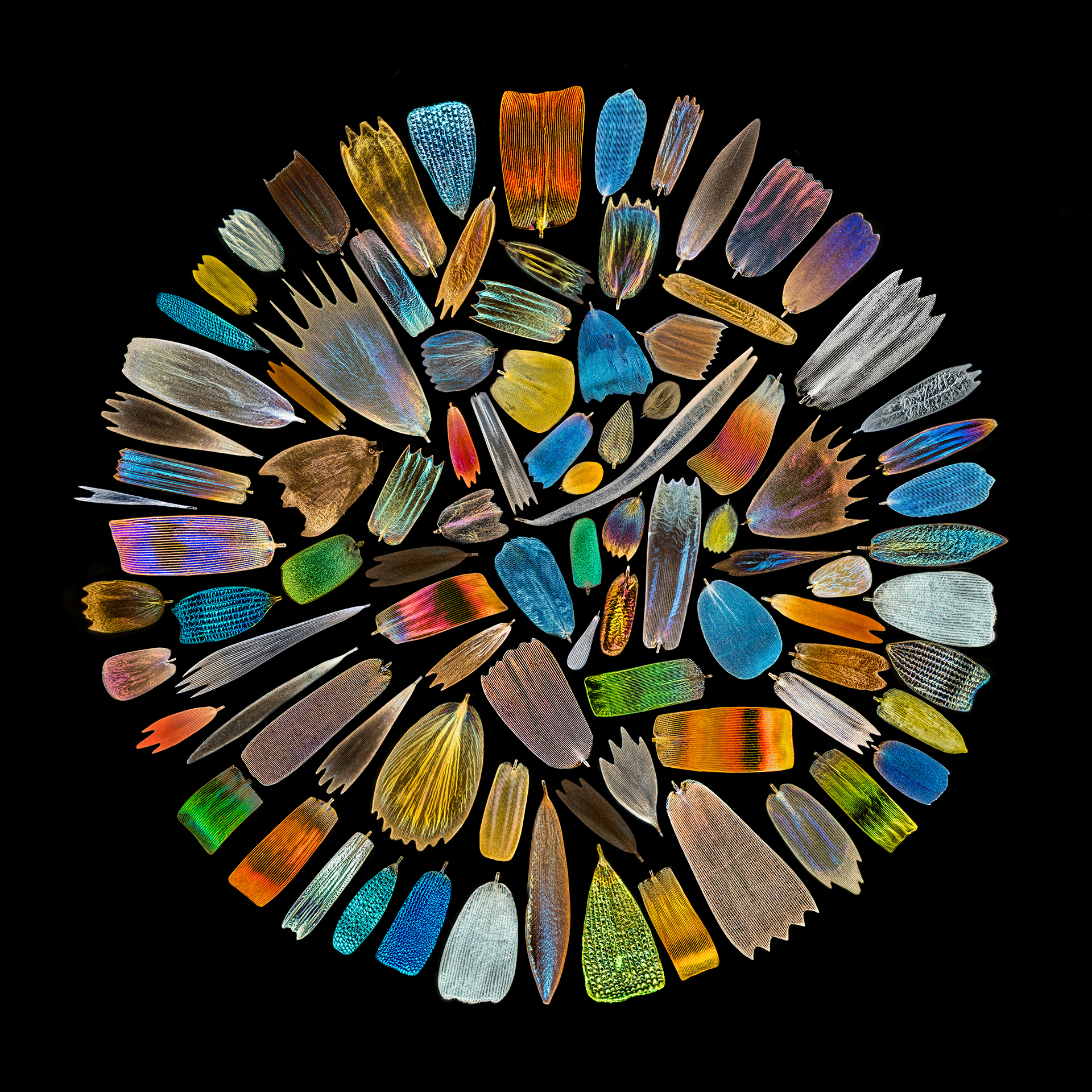 Asia-Pacific regional prize was awarded to XinPei Zhang (China). Scales collected from the wings of over 40 species of butterflies were photographed individually and finally assembled into this image. |
Honorable Mentions
 Anther of Arabidopsis arenosa stained with aniline blue, Z-stack of confocal sections (maximum intensity projection). | 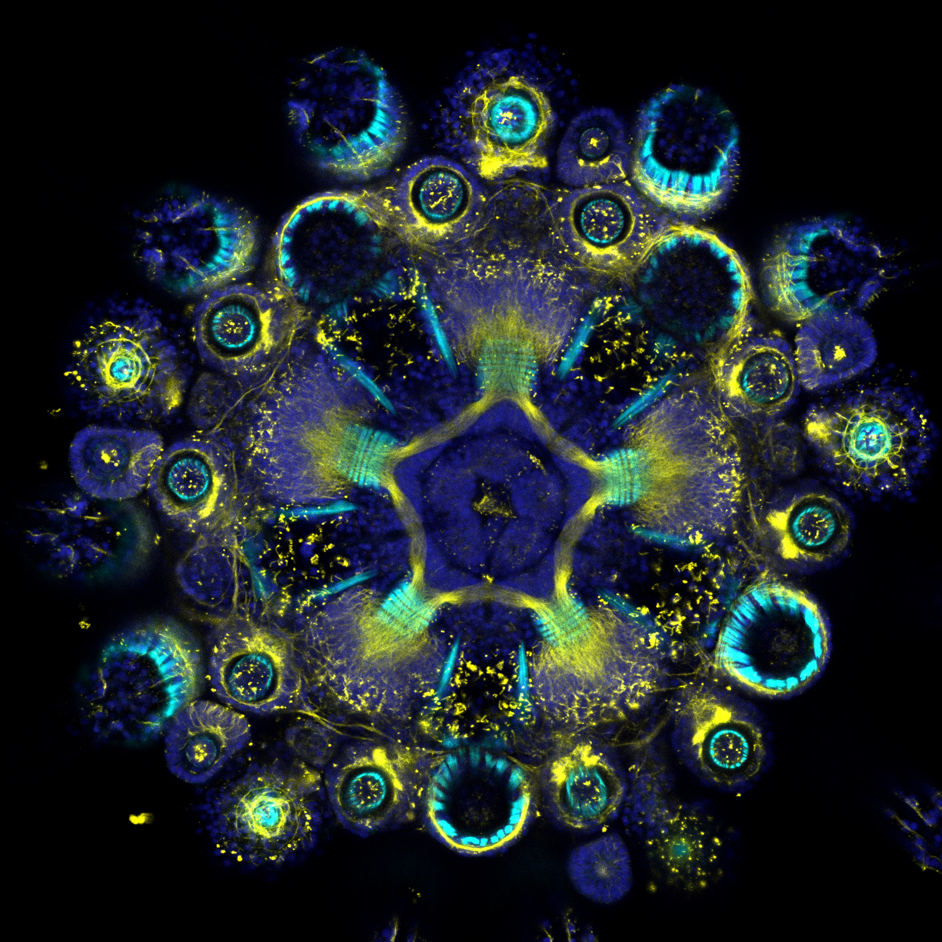 Whole-mount immunofluorescence showing the muscles (cyan, F-actin) and the nervous system (yellows, acetylated tubulin) counterstained by the nuclei (blue, DNA) of a clarified and decalcified post-metamorphic sea urchin juvenile (Paracentrotus lividus). | 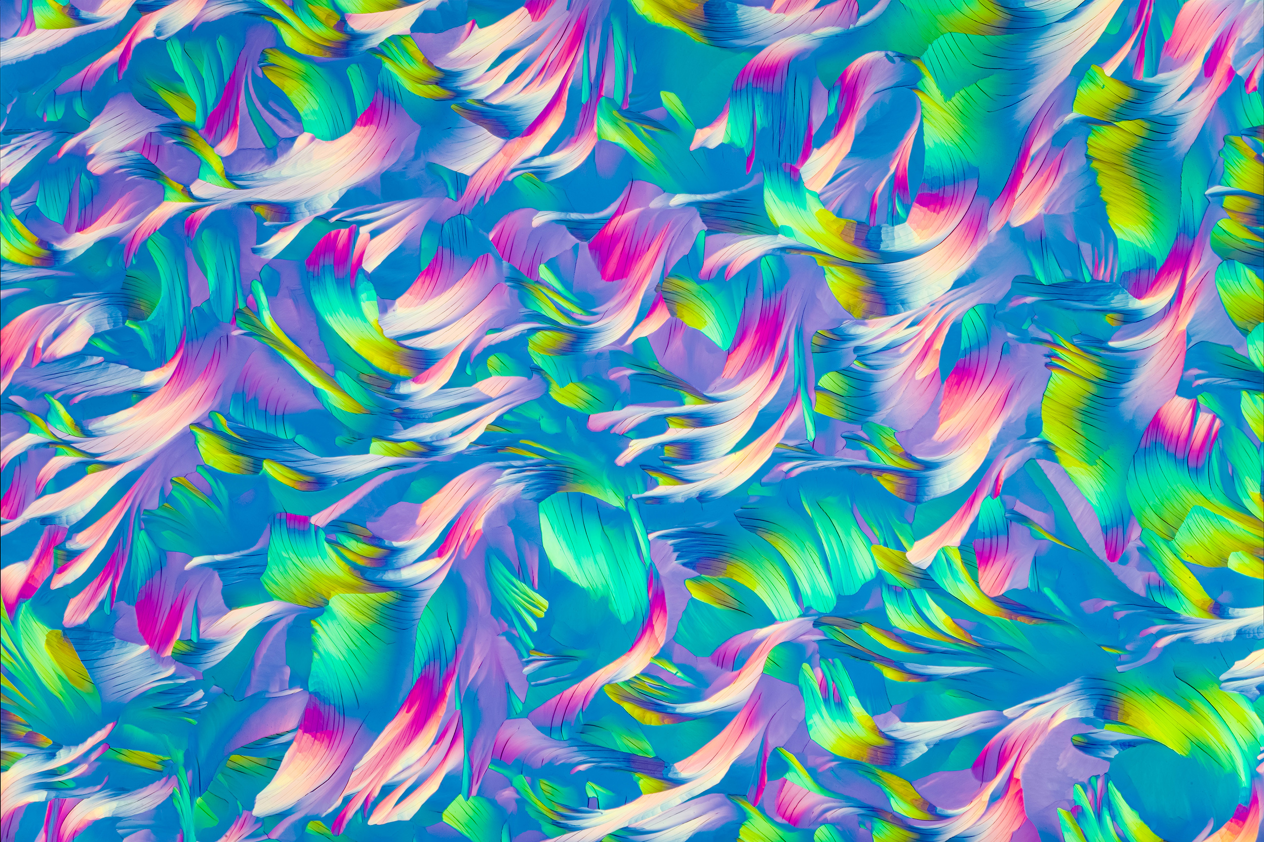 A polarized light micrograph of crystals formed by the rapid evaporation of water from an aqueous solution of Beta-Alanine (a popular training supplement), and l-Glutamine (an essential amino acid). Home-made tunable wave plate. |
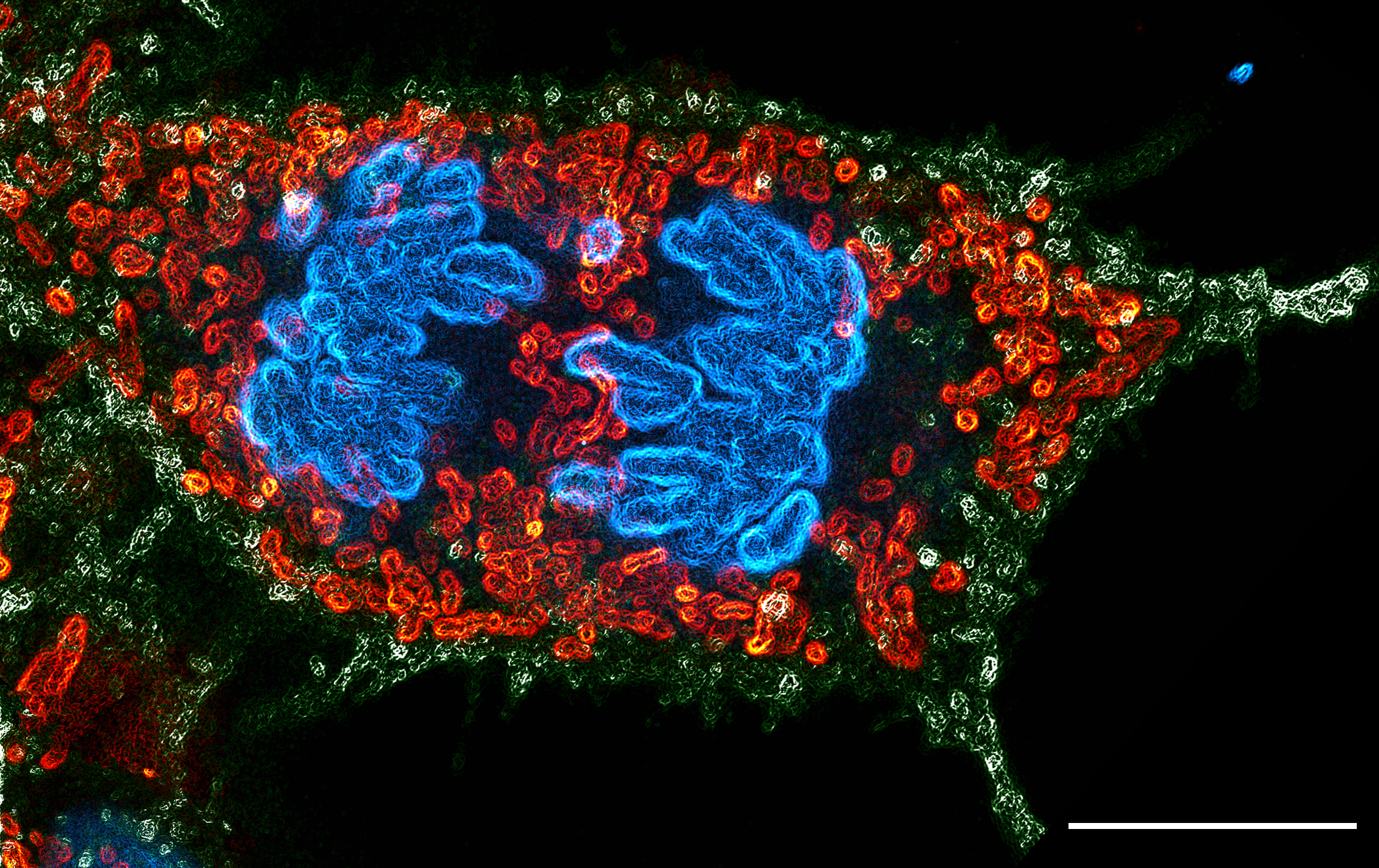 The sample shows a dividing human embryonic kidney (HEK) 293 cell. Green marks the cellular boundary, red marks the mitochondria, while blue shows the separating chromosomes. | 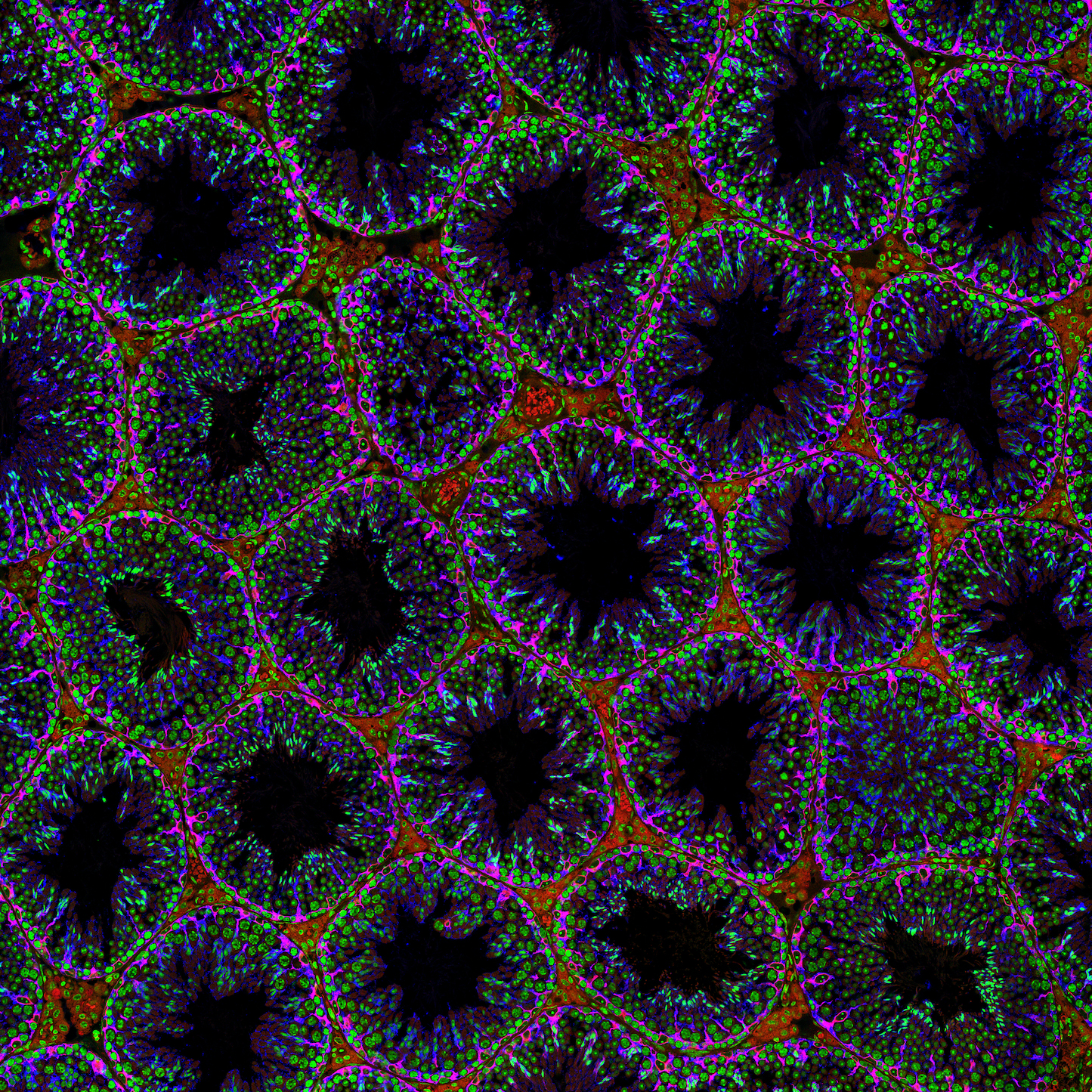 This immunofluorescence image of an adult mouse testes section was stained for beta III tubulin (blue) and vimentin (red), two markers specific to Sertoli cells. In green are nuclei stained with DAPI, and the various stages of sperm can be distinguished by their nuclear morphology. |  “Pollock’s Glia.” 3D reconstructed immunostaining image of astrocyte (GFAP+, white), oligodendrocyte (NG2+, blue), and microglia (IBA1+, red) in the brain white matter, which appears highly similar to Pollock’s abstract paintings. |
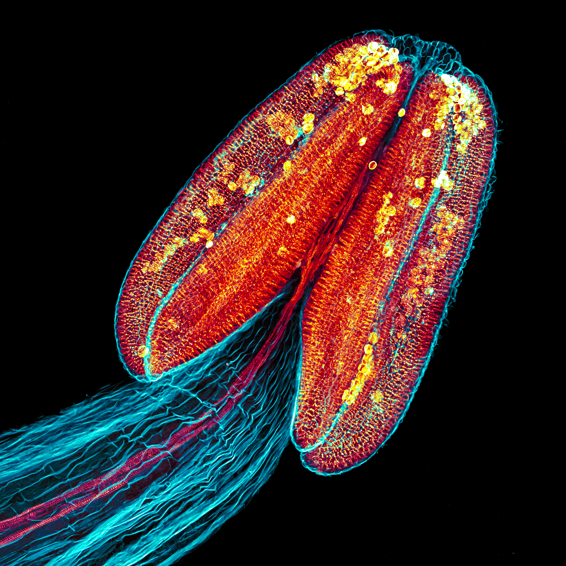
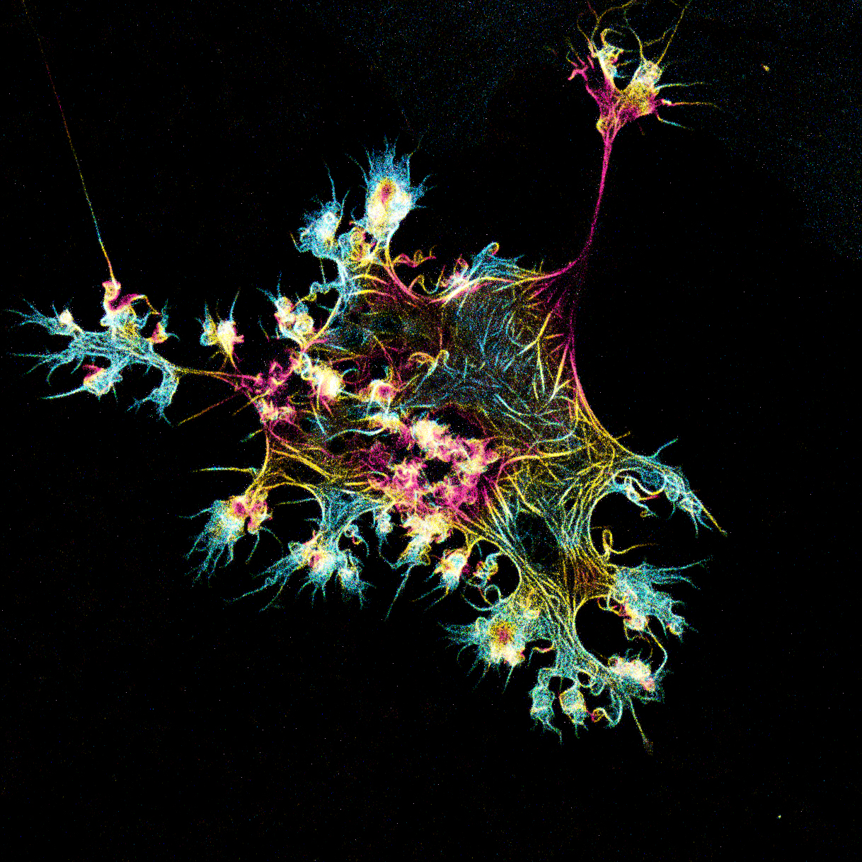 Microfilament structure in U2OS labeled with fluorescent protein. |  Cultured neurons are a good model to test enzyme replacement therapy (ERT) for many neurological disorders. In this experiment Efimova looked at uptake of engineered enzyme in rat cortical neurons (staining of enzyme is not shown). MAP2 staining was added to visualize outlines of neurons. | 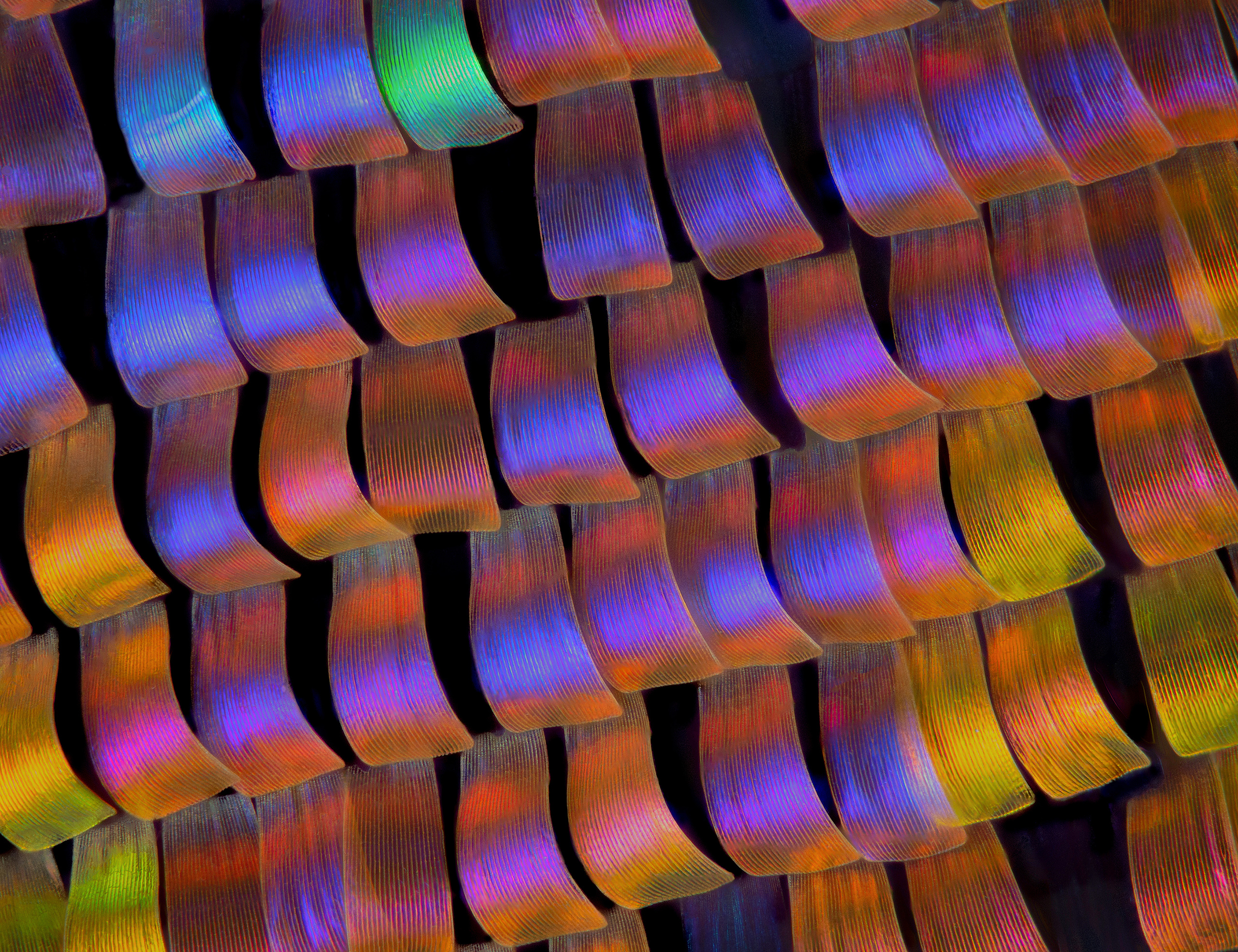 This image captures wing scales of the twilight moth Urania ripheus. The image was achieved using the focus stacking technique, for which many photos are taken at different depths and then stacked using software. |
Download Wallpapers
Download the Image of the Year Award 2020 wallpaper package now for free and beautify your screen!
Download the wallpaper package for desktop (ZIP, 40.2 MB)
Download the wallpaper package for mobile (ZIP, 26.8 MB)
Virtual Backgrounds
Download your favorite virtual backgrounds and add them to your meetings! Download the virtual backgrounds package for your meeting application (ZIP, 3.36 MB) |  |
Sorry, this page is not
available in your country.
Sorry, this page is not
available in your country.