Not Available in Your Country
Sorry, this page is not
available in your country.
Overview
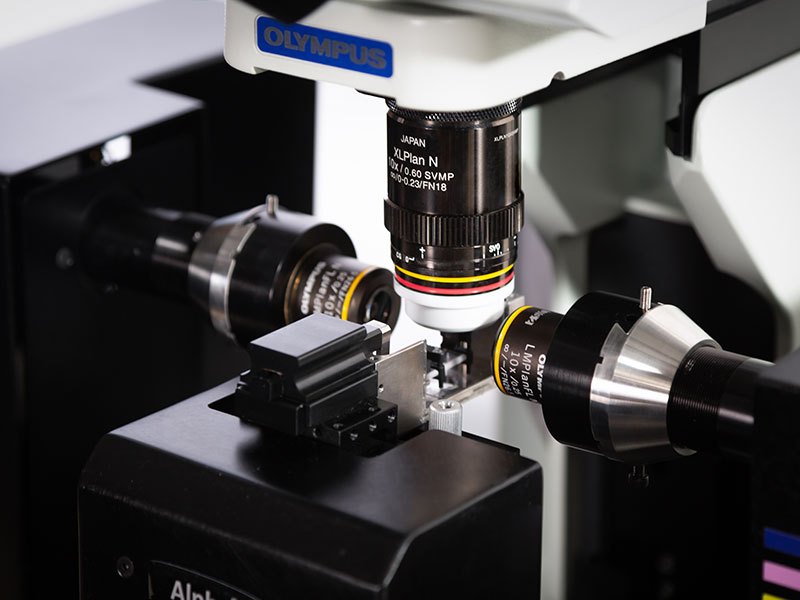
 | Flexibilité et performances de qualitéLe système Alpha3 de PhaseView a été conçu pour offrir aux chercheurs une solution à feuillet de lumière flexible et très performante. Le microscope à fluorescence Alpha3 offre une focalisation du balayage optique intelligente pour une imagerie uniforme et sans artefacts à travers l’ensemble du champ de vision. Le système peut être facilement mis à niveau avec des options telles que le balayage rapide (balayage 3D intelligent), le système de quadrillage XY et le contrôle de la température, afin de répondre à divers besoins en matière d’expérimentation. Le logiciel QtSPIM fournit une interface claire et intuitive permettant de collecter des images X, Y, Z, θ, T et λ à une vitesse maximale. |
|---|
Quatre valeurs clés du système Alpha3
|    |
Caractéristiques techniquesVeuillez visiter le site Web de PhaseView pour obtenir les caractéristiques techniques du produit. En savoir plus sur les caractéristiques techniques du système Alpha3 |
Besoin d’aide ? |
Applied Technologies
Double éclairageGrâce à l’intégration d’unités de double éclairage, le microscope à feuillet de lumière multidirectionnel produit des images sans artefact des échantillons qui absorbent ou diffusent la lumière. Couplé au module de focalisation du balayage optique, le système Alpha3 assure une netteté optimisée à travers l’ensemble du champ de vision pour une qualité d’image incomparable. | 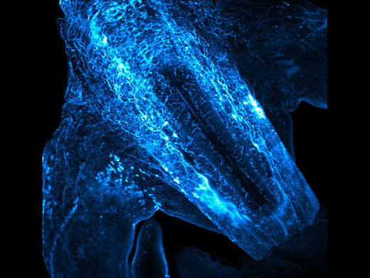 Embryon de souris |
Focalisation du balayage optique en temps réelLa focalisation du balayage optique en temps réel pallie les contraintes de résolution spatiale et temporelle liées à l’acquisition d’images 3D. Ce module en option maintient la focalisation du feuillet de lumière à travers l’ensemble du champ de vision pour assurer une clarté optimale de l’image. En d’autres termes, le champ exposé se trouve toujours dans la région la plus mince du feuillet de lumière. Ce principe repose sur une lentille réglable assurant le balayage du plan focal de façon synchronisée avec l’obturateur roulant de la caméra. 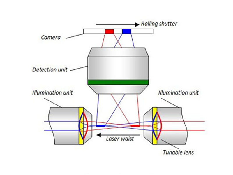 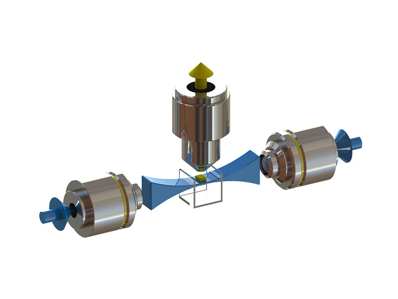  Cleared mouse brain image taken without (left) and with sharp optical sectioning (right). |
Balayage 3D intelligentAu lieu de déplacer l’échantillon pour effectuer un balayage dans le plan Z, il est placé dans une position fixe tandis que les plans d’éclairage et de détection se déplacent simultanément à travers l’échantillon. Comme l’échantillon demeure constamment en position stable, les vibrations et les perturbations sont atténuées. Le balayage du plan d’éclairage est assuré à l’aide d’un miroir galvanométrique, tandis que le balayage du plan de détection est réalisé à l’aide d’un dispositif de focalisation à distance pour produire une imagerie 3D sans perturbation à une vitesse élevée de 75 images/seconde. | 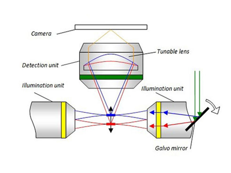 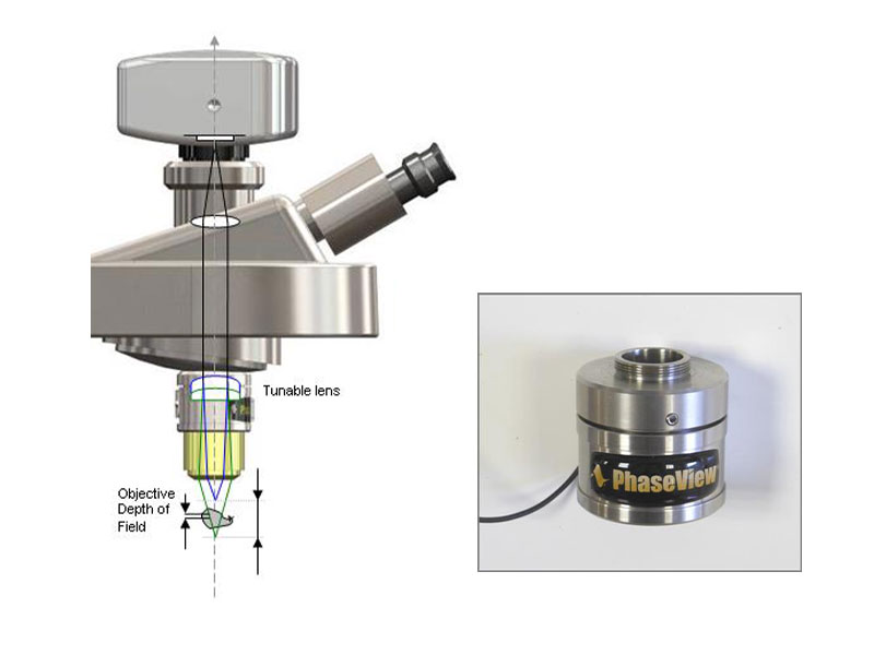 |
 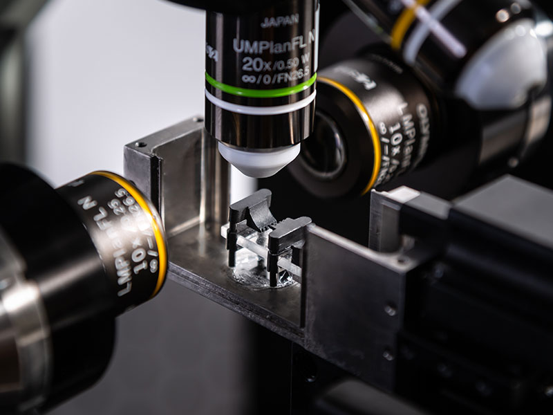 | Conception polyvalenteDivers porte-échantillons et accessoires de montage permettent d’acquérir des images de petits échantillons comme d’organes entiers. Pour l’imagerie in vivo, des moyens de contrôle précis de la température et du taux de CO2 sont également disponibles. |
|---|
Interface utilisateur intelligenteLe logiciel QtSPIM offre une interface intuitive et clairement présentée permettant la collecte d’images 3D selon une procédure efficace.
| 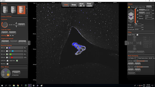 Interface utilisateur QTSPIM d’imagerie 3D. |
Besoin d’aide ? |
Application Gallery
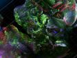
 Projection d’intensité maximale d’embryons de souris, transparisation iDisco | 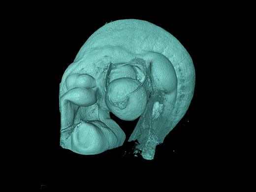 Volume 3D d’embryons de souris, transparisation iDisco |
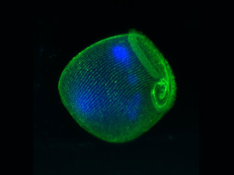
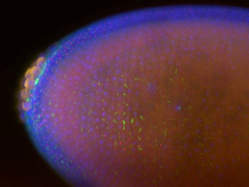
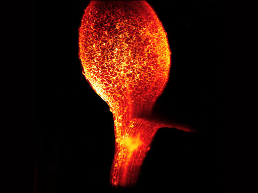 Marquage in vivo de la membrane des cellules d’une feuille d’Arabidopsis à 10x | 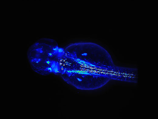 Fluorescence RFP et GCamp des neurones moteurs de larves de poissons-zèbres vivantes, reproduction avec l’aimable autorisation de BioEmergences, France-BioImaging |
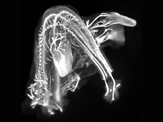
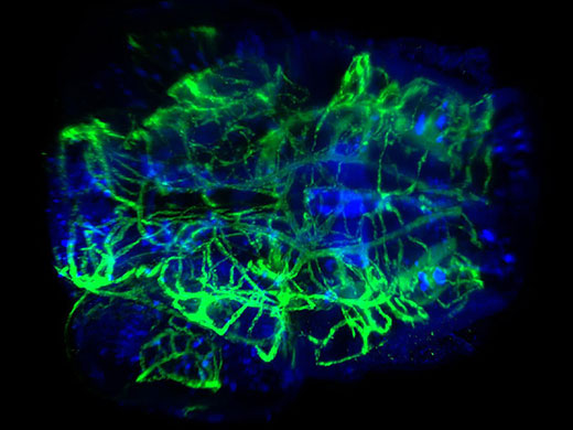 Tête transparisée d’une larve de poisson-zèbre colorée pour observer le système vasculaire et neural, grossissement 20x | 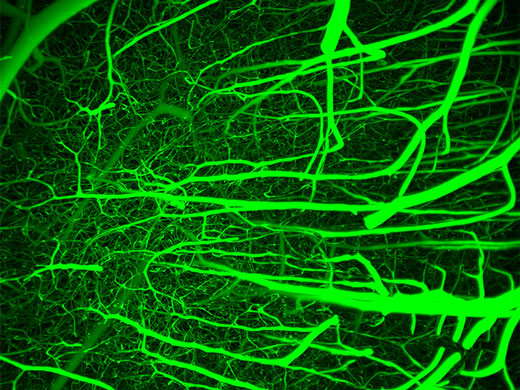 Système vasculaire du cerveau d’une souris, reproduction avec l’aimable autorisation des Dr. Beth Friedman et Hannah Liechty, Kleinfield Laboratory UC San Diego. |