Regenerative medicine harnesses cutting-edge technologies such as stem cell therapy and tissue engineering to repair the body, presenting innovative solutions for medical conditions from injuries to degenerative diseases. In this field, microscopy plays a pivotal role in every stage of your research workflow. Whether you are culturing stem cells, monitoring engineered tissues, or conducting in-depth cellular analyses, our microscopy solutions are tailored to elevate your research. Enhance
standardization and efficiency during cell culture with our incubation monitoring system. Effortlessly capture expert-quality brightfield and fluorescent images with our all-in-one digital imaging system. For high-speed, precision analysis of cellular and tissue dynamics, explore our range of laser scanning and spinning-disk confocal systems.
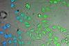
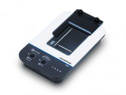
Remotely monitor, analyze, and share your cell cultures’ health, cell count, and confluency using the reliable quantitative data provided by the automated CM30 incubation monitoring system. The system enables label-free observation, reduces the risk of damage to your cultures, and standardizes your culture workflow.
Automatically collects quantitative data on the health and confluency of your cultures
Monitor, analyze, and share your cultures' progress remotely from a PC or tablet
Equipped with oblique epi-illumination for label-free observation
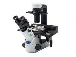
The compact, ergonomic CKX53 inverted microscope’s simple phase contrast system enables simple, high-contrast cell culture observation, accommodating a variety of containers. Its inversion contrast (IVC) technique offers clear pseudo-3D observation, easing workflows including live cell imaging, cell sampling and handling, and fluorescence.