Monitoring Spheroid Formation in U-Bottom Multiwell Plates Using an Incubation Monitoring System
Experimental Outline
Spheroids are 3D cell aggregates designed to mimic features of tissue in vivo, making them valuable for a range of assessments such as testing the differentiation potential of stem cells or drug efficacy. U-bottom plates are often used for spheroid formation. This experiment was conducted to evaluate the Olympus Provi™ CM20 incubation monitoring system’s benefits for monitoring spheroid formation on U-bottom plates.
Experimental Procedure
Mouse neural stem cells were seeded on a 96-well U-bottom plate (Sumitomo Bakelite, Cat. No. MS-9096U) at a density of 2,500 cells/0.1 mL/well using Knock Out DMEM/F-12 supplemented with 1x B27, 1x GlutaMax, 20 ng/mL bFGF, 10 ng/mL EGF, 0.0002% heparin, 1% penicillin-streptomycin.
The prepared 96-well plate was placed on the Provi CM20 incubation monitoring system. The focus position was set manually, and the cells were imaged automatically by the CM20 system every hour for a period of 60 hours to monitor spheroid formation.
Results
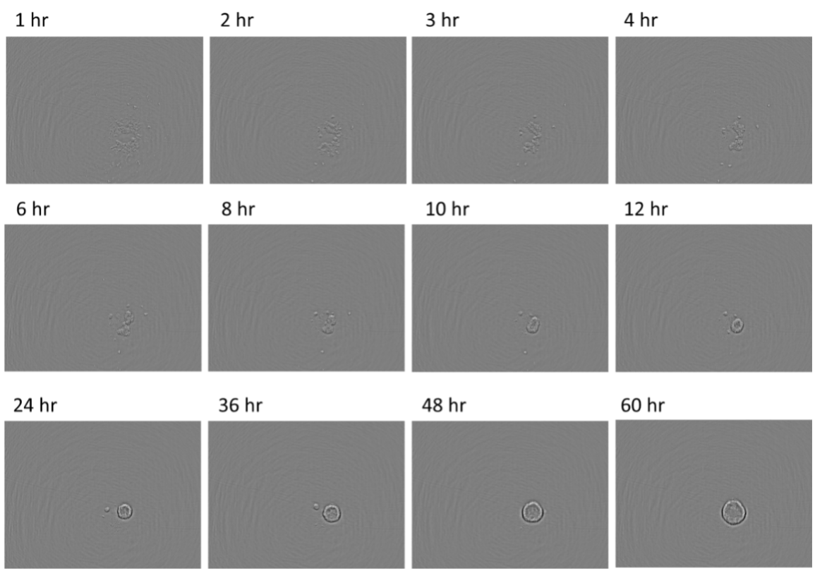
Figure 1. Spheroid formation from mouse neural stem cells
By assessing the acquired images, we were able to conclude that after seeding, the cells came together rapidly, became spheroids in approximately 10 h, and then gradually increased in size.
Benefits of Cell Culture Assessment Using the CM20 System
Imaging of with the Provi CM20 system was successfully performed using U-bottom plates. A major advantage of this method is that the cells can remain undisturbed in the incubator, mitigating the imaging process's impact on spheroid formation. This experiment also demonstrates that with the CM20, the focus position only needs to be set up once, and then can be maintained for multiple days, even for objects in suspension. Based on these results, we can conclude that the Provi CM20 system can be used to perform high-throughput time-lapse analysis using U-bottom 96-well plates. This is especially useful for drug screening in spheroid tumor models. We also predict that the CM20 system could be used to monitor the differentiation of stem cells (for example, neural stem cells) or embryoid bodies (EB), and we intend to test this method for such applications in future experiments.
Acknowledgments
This application note was prepared with the help of the following researcher:
Takahiro Yamaguchi, PhD, Principal Researcher, ACEL, Inc.
Evident, the Evident logo, and Olympus Provi are trademarks of Evident Corporation or its subsidiaries.
Products Related to This Application
Sorry, this page is not
available in your country.