Remotely Monitoring the Primary Culture of Bone Marrow-Derived Mesenchymal Stem Cells
Experimental outline
The bone marrow-derived mesenchymal stem cell is multipotent. Therefore, it is the target of active research and clinical applications as the source of cells for regenerative medicine. When bone marrow fluids or bone marrow cells are seeded on Petri dishes, the mesenchymal stem cells proliferate as adherent cells. As a result, the mesenchymal stem cells can be separated from blood cells that are floating. Here we report the remote monitoring results of the primary culture of mesenchymal stem cells using the Olympus Provi™ CM20 incubation monitoring system.
Experimental procedures
Commercially available human bone marrow cells were seeded on a 24-well plate (Sumitomo Bakelite, Cat. No. MS-80240) at the density of 950,000 cells/0.5 mL/well using Iscove’s Modified Dulbecco’s Medium (IMDM) supplemented with 10% fetal bovine serum (FBS) and 1% Penicillin-Streptomycin. Cells were imaged using the CM20 system every one hour with autofocus. Since the proliferation of adherent cells was observed on day 4 of culture, they were monitored for up to 6 days after exchanging media.
Results
Just after seeding
| Day 4 (before media exchange)
| Day 4 (after media exchange)
| Day 6
|
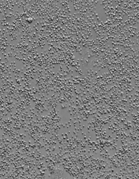
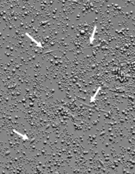
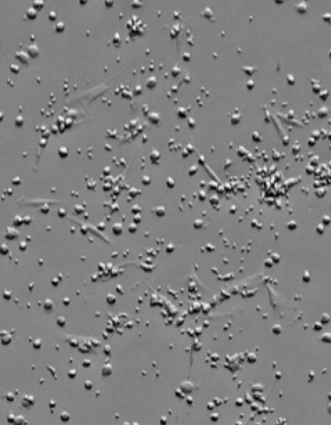
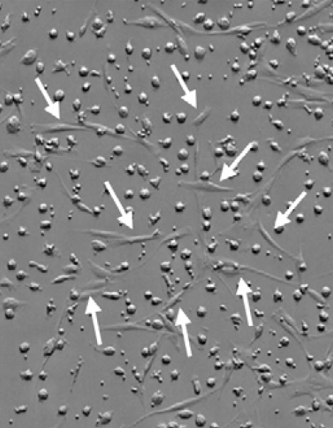
Figure 1. Primary culture of bone marrow-derived mesenchymal stem cells. Arrows indicate bone marrow-derived mesenchymal stem cells.
Most of the cells were blood cells, and no mesenchymal stem cells were observed immediately after seeding. However, on day 4 after seeding, mesenchymal stem cells with the morphology of a spindle body appeared. Subsequently, the media was exchanged to remove the blood cells. On day 6 after seeding, the mesenchymal stem cells were found to proliferate into a large number of cells.
Benefits of using incubation monitoring system assessments
Primary culture of bone marrow-derived mesenchymal stem cells should be conducted as gently as possible until adherent cells appear. Shaking, which could occur during microscopic observation or medium change, should be avoided as much as possible. Here we showed that the CM20 system enabled us to remotely monitor the cells under incubation. By using the CM20 system, we shortened the operation time and could proceed to the rest of the experiment at the proper time.
Acknowledgments
This application note was prepared with the help of the following researcher:
Takahiro Yamaguchi, PhD, Principal Researcher, ACEL, Inc.
Products Related to This Application
Sorry, this page is not
available in your country.