Not Available in Your Country
Sorry, this page is not
available in your country.
- Overview
- Applied Technologies
- AI Solutions for Confocal Microscopy
- Configurations
- Specifications
- Resources
Overview
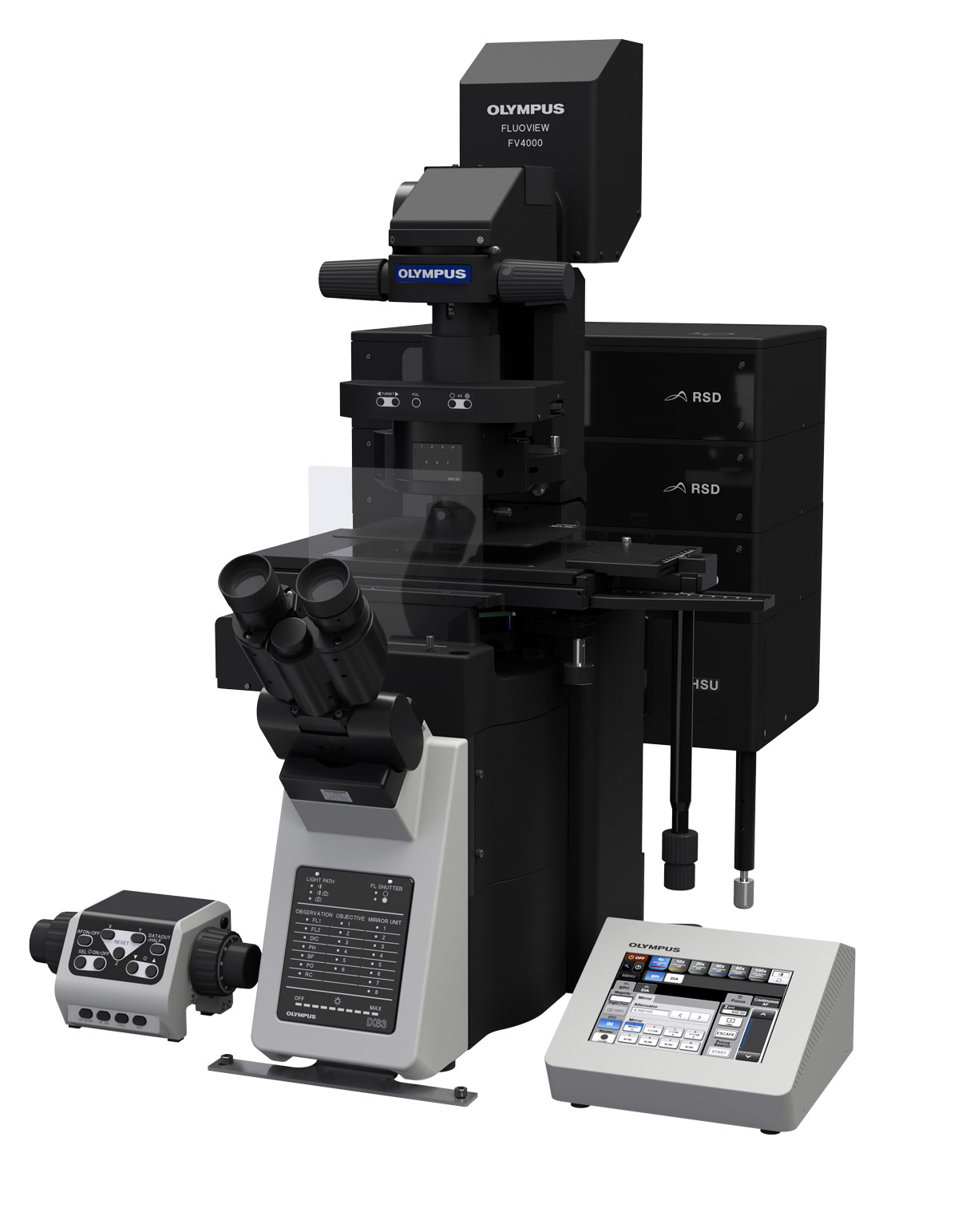
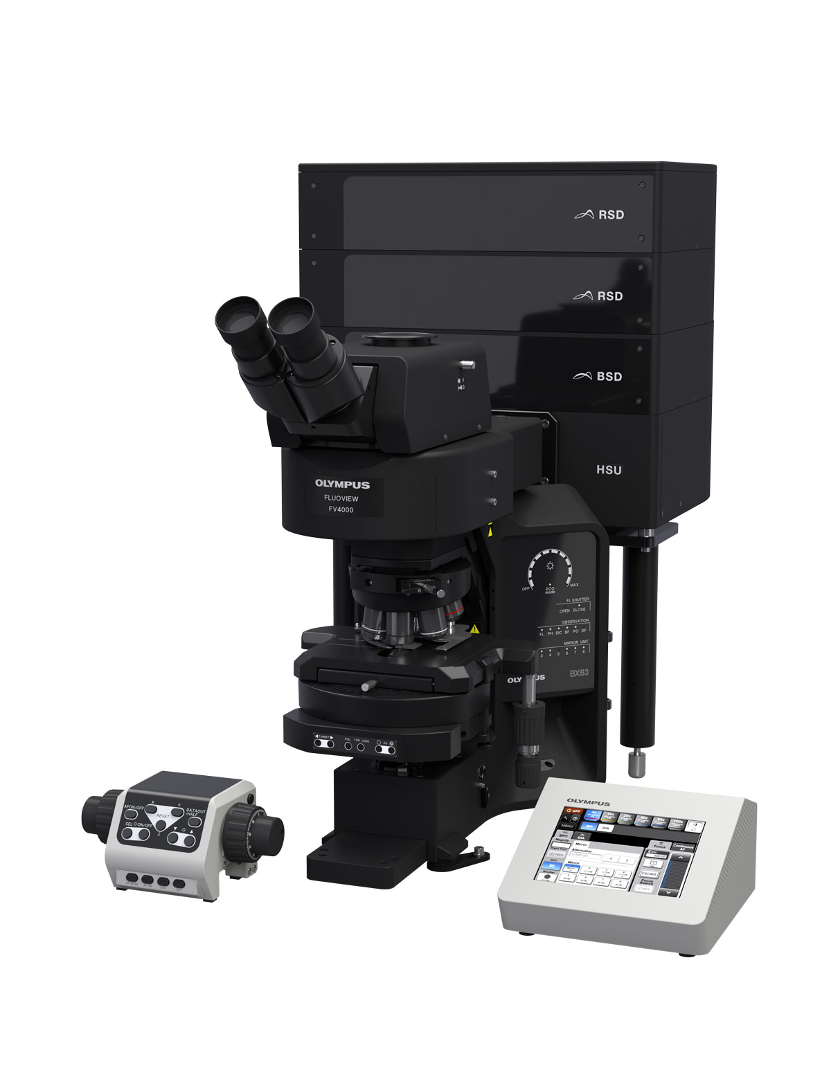
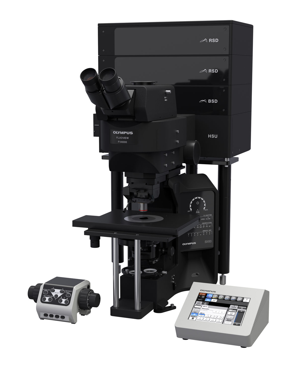
 | Transforming Precision ImagingEmpower Your Confocal Microscopy Imaging ExperimentsTransform your images with the new FLUOVIEW™ FV4000 confocal laser scanning microscope. Advanced imaging technology enables higher precision images to empower researchers with more reliable data from their samples. With our breakthrough SilVIR™ detector at the core of the system, achieve much lower noise, higher sensitivity, and improved photon resolving capabilities. With the FV4000 confocal microscope, researchers can acquire higher-quality, quantitative image data in less time and with less effort. |
|---|
Experience the systems innovations, including:
*As of October 2023. |
Easy-to-Acquire, Quantitative Confocal DataThe FV4000 confocal microscope uses our advanced, silicon-based SilVIR™ detector that makes it easier than ever to acquire precise, reproducible data. SilVIR Next-Generation Detector Technology The SilVIR detector combines two advanced technologies—a silicon photomultiplier (SiPM) and our patented* fast signal processing design.
*Patent number US11237047 Learn more about the SilVIR detector |
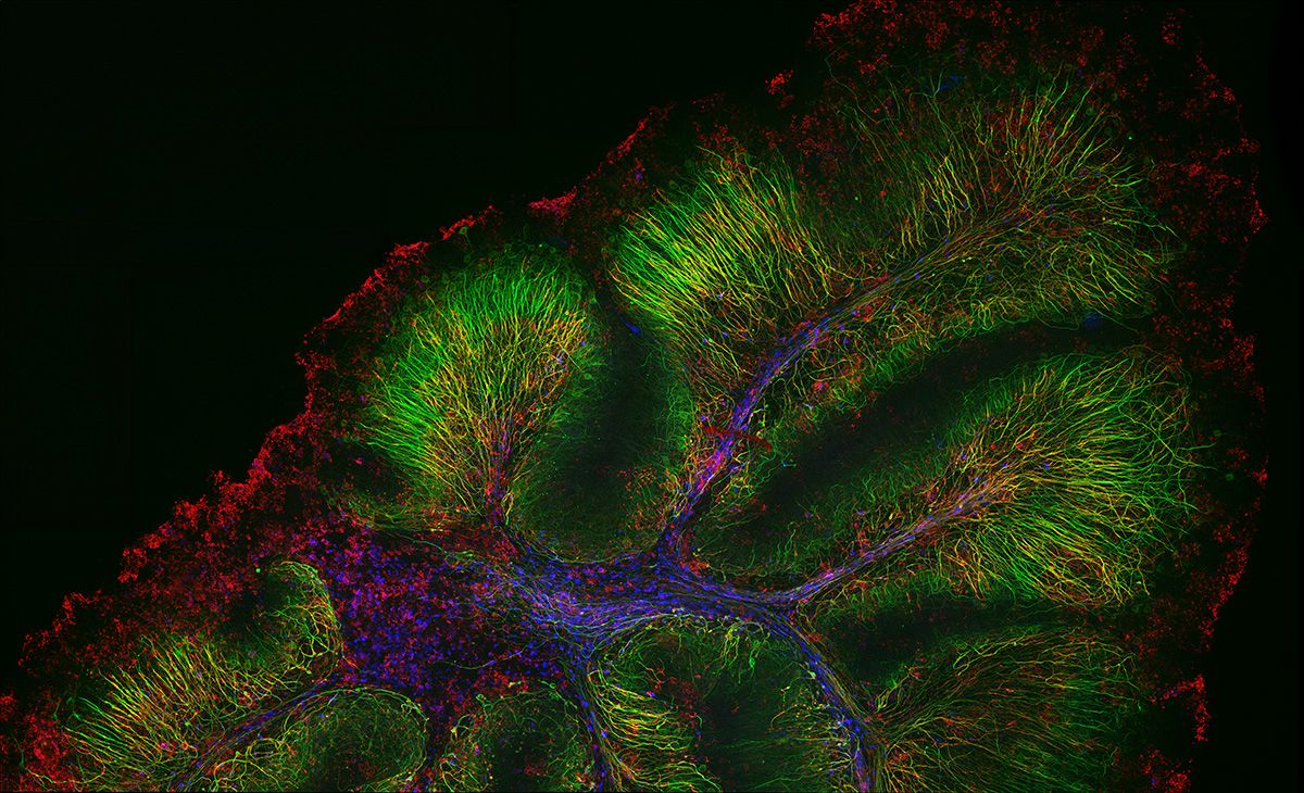
Neurofilament-heavy chain (NFH) in green, myelin basic protein (MBP) in red, glutathione S-transferase pi 1 (GSTpi) in blue. Mouse cerebellum captured with a UPLXAPO40X objective.
|
|
|
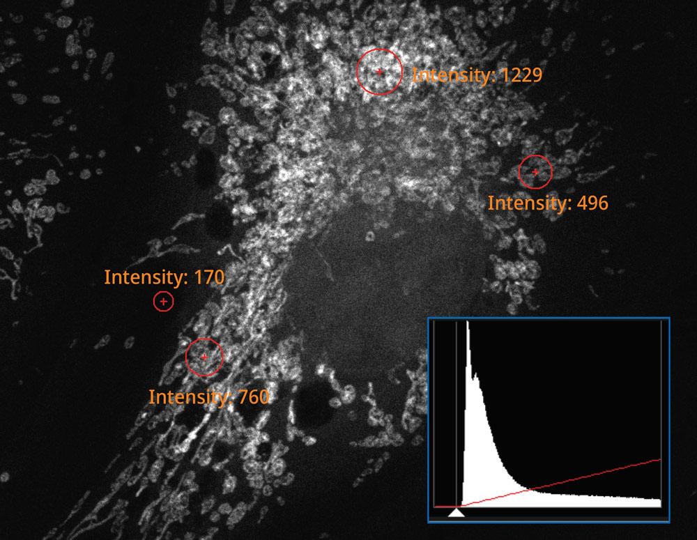
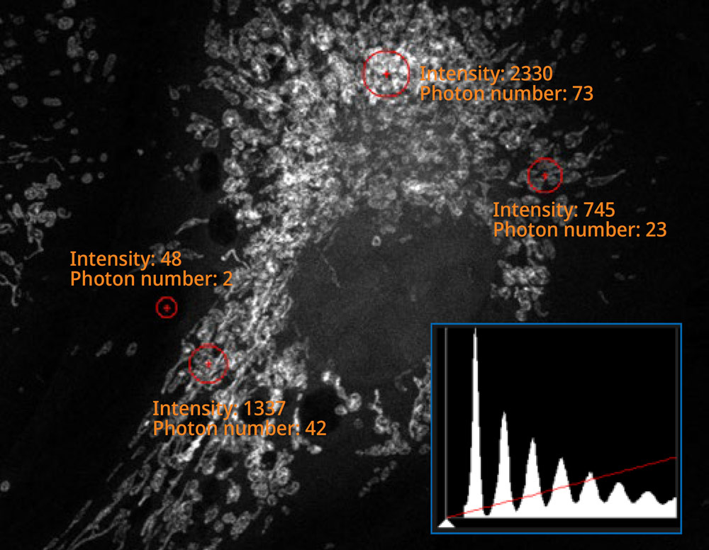
The histogram on the image captured using the SilVIR detector shows a discrete pattern where the intensity can be converted to the photon number. The detector’s fluorescence intensity can be quantified as the photon number, and the background level is extremely low. |
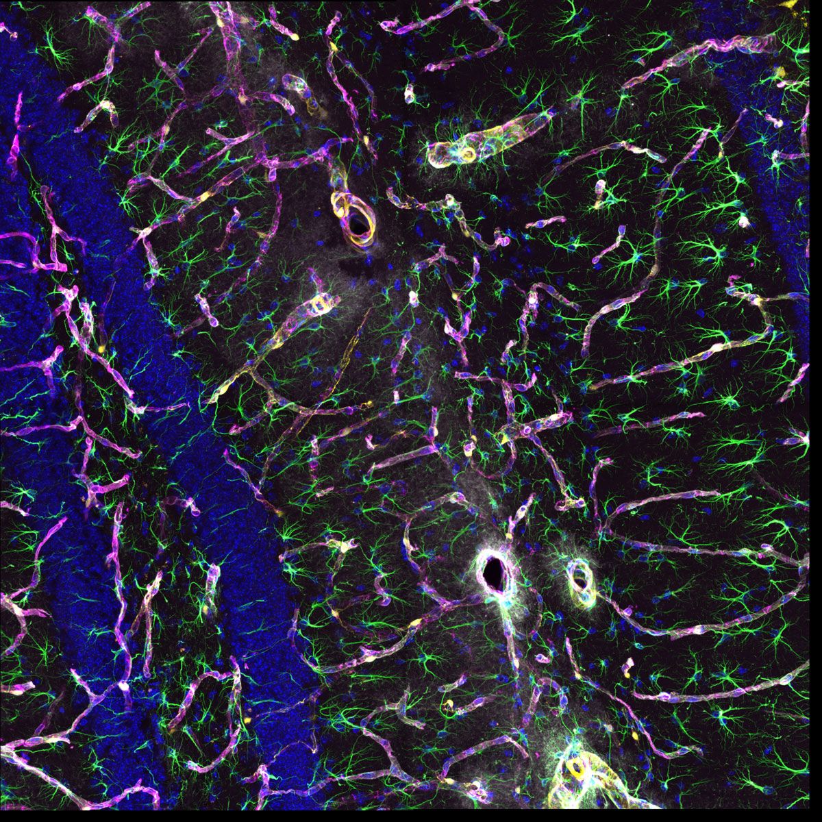
More Information from Your Confocal ImagesThe system’s updated TruSpectral technology combined with high sensitivity SilVIR detectors enable you to see more by making it possible to multiplex up to six channels simultaneously.
|
Flexible Macro to Micro ImagingThe macro-to-micro workflow enables you to easily observe the target sample from the macro level—whole body or tissue—down to the cell or subcellular level.
| ||||
Gentler High-Speed Time-Lapse Confocal Imaging | |
Related VideosHeLa cells labeled by MitoView 720. XYZT imaging by 1K resonant scanner for 30 min. | Time-lapse imaging is easier with smart features:
|
Reproducible Image Data Between Users and SystemsThe SilVIR detector has less sensitivity loss over time compared to previous-generation detector technologies. With our laser power monitor (LPM) and TruFocus™ Z-drift compensator, achieve reproducible images under consistent conditions for better reproducibility. Different users on different days can acquire the same precise images using the same settings. Even the images acquired by different FV4000 microscopes can be compared and discussed using the same photon number intensity scale. |
Microscope Support and Service You Can Count OnWe designed the FV4000 system to be easy to maintain:
We stand behind our products with a commitment to fast service and technical support. We offer various support plans to keep your microscope running at peak performance at a predictable cost as well as remote support options, so you don’t need to wait for an engineer or specialist to visit if you’re having an issue.
|
Need assistance? |
Applied Technologies
See Further with NIR-Enabled Confocal MicroscopyThe system’s enhanced technologies provided expanded multiplexing to see more in one image. NIR imaging offers greater multiplexing capabilities by extending the excitation (λ_Ex) and detection (λ_Em) spectral profile of the FV4000 system. This enables additional dyes to be used to help minimize emission signal overlapping.
|
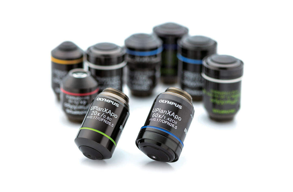
High-Quality Optics for Efficient NIR Fluorescence ImagingThe FV4000 system's optical elements have a high transmission from 400 nm to 1300 nm, including the galvanometer and resonant scanner, which are coated in silver rather than aluminum. Our award-winning X Line™ objectives are corrected for chromatic aberrations between 400–1000 nm. They also have a higher numerical aperture, excellent flatness, and very high transmittance from UV to NIR, increasing the multiplexing capabilities. For improved colocalization reliability, our specialized A Line™ (PLAPON60XOSC2) oil immersion objective (ne~1.40) significantly minimizes chromatic aberration for strict colocalization analysis. |
|
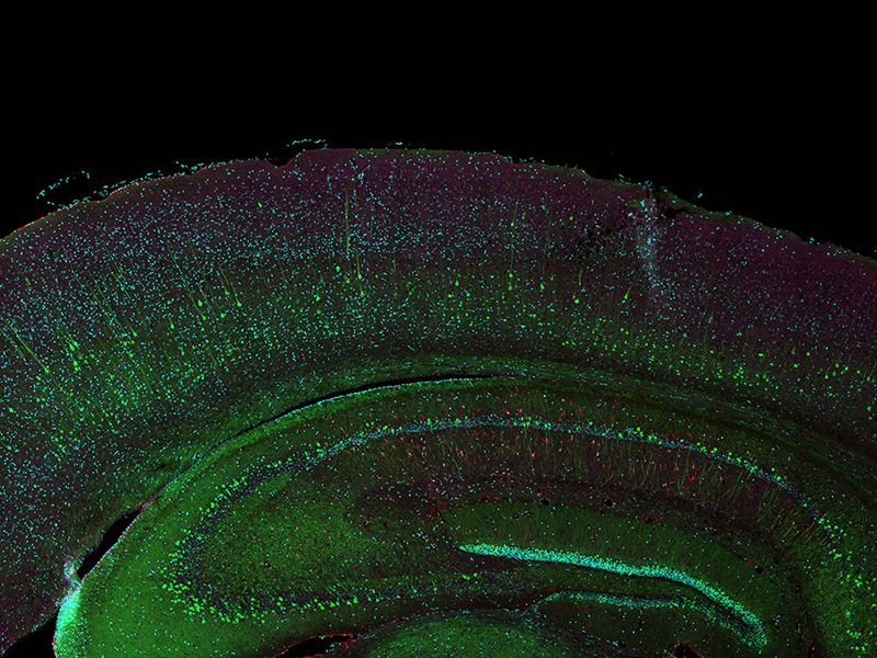  A total of 77 four-channel XYZ positions (11 × 7) were acquired using a 1K resonant scanner within 16 minutes to create the stitched image, which used to require 2 hours using a galvanometer scanner. The coronal section of an H-line mouse brain, cyan; DAPI (cell nuclei), green; YFP (neuron), yellow; Cy3 astrocytes, magenta; AlexaFluor 750 (microtubule). Sample courtesy of: Takako Kogure and Atsushi Miyawaki, Cell Function Dynamics, RIKEN CBS. | High-Quality Confocal Images at High SpeedA unique combination of advanced technologies delivers high-quality images faster than conventional laser scanning microscope systems.
|
|---|
Simple, Precise Super Resolution ImagingCapture super resolution images using the FV4000 microscope with no dedicated hardware.
|  Confocal mode 1AU (left) versus super resolution mode (right) |
High-Resolution 3D Images in Thick Samples | |
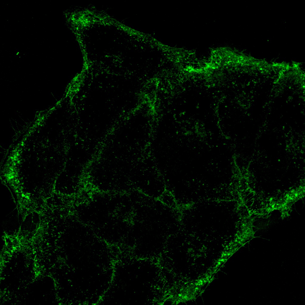
Related VideosHeLa cell spheroid labeled by DAPI (cyan, cell nuclei) and AlexaFluor790 (magenta, Ki-67). Imaging of the spheroid’s whole volume was possible by NIR 785 nm, although only surface area cell nuclei observation was possible using a 405 nm laser. | When imaging thicker samples, the FV4000 microscope enables you to capture high-resolution, 3D images.
|
Precise Dynamics of Live Cells with Less Damage
|
Clear Images at DepthUse our silicone immersion objectives with the FV4000 microscope and achieve clear images of features and structures deep within your sample. Silicone oil has a refractive index close to that of live cells or tissue, greatly reducing the spherical aberration as compared to air, water, or other oils. With less aberration, you can achieve clearer images of your sample at depth. And silicone immersion oil does not dry out at 37 ℃ (98.6 °F), making it effective for long-term time-lapse imaging. | Related Videos |
Need assistance? |
AI Solutions for Confocal Microscopy
Take your confocal imaging to the next level and save time during data analysis. While the microscope’s signal-to-noise ratio is already very good, TruAI™ technology denoise can further reduce the noise for stunning, data-rich resonant images. To speed up image analysis, you can pretrain an AI model so that the system can automatically segment your image data, greatly reducing the workload of this often time-consuming manual process. Then, TruAI technology further streamlines the analysis so that you can get your data quickly. |
TruAI Noise ReductionImprove your resonant scanner image quality by incorporating TruAI noise reduction. Although resonant scanner images are effective in capturing cellular dynamics at high speeds with low damage, this usually causes a compromise in the S/N ratio. TruAI noise reduction can improve these images without sacrificing time resolution using pre-trained neural networks based on the noise pattern of the SilVIR™ detectors. These pre-trained TruAI noise reduction algorithms can be used for on-the-fly processing as well as post processing. | |
 Processed with TruAI noise reduction (right) Brain sample: coronal section (50 μm) of a mouse brain stained with DAPI (nuclei, cyan), GFAP (astrocytes, green/488), MAP2 (microtubule-associated protein 2, neurons, and dendritic processes, cyan/647) and MBP (myelin basic protein, red/568). Sample courtesy of: Sample preparation Alexia Ferrand; sample acquisition Sara R. Roig and Alexia Ferrand. Imaging Core Facility, Biozentrum, University of Basel. |  Processed with TruAI noise reduction (right) HeLa cell mitochondria labeled by MitoView 720 acquired using a 1K resonant scanner. The maximum photon number was 3 photons. |
TruAI Image SegmentationImage analysis requires data extraction using segmentation techniques based on intensity value thresholds. However, this can be time-consuming and is affected by the sample conditions. TruAI image segmentation using deep learning helps streamline image processing and minimize sample variables for more accurate image analysis. It enables you to segment very weak fluorescence images or tissues that are usually difficult to extract using the simple thresholding method. |  TruAI detects the glomeruli features (right) |
Need assistance? |
Configurations
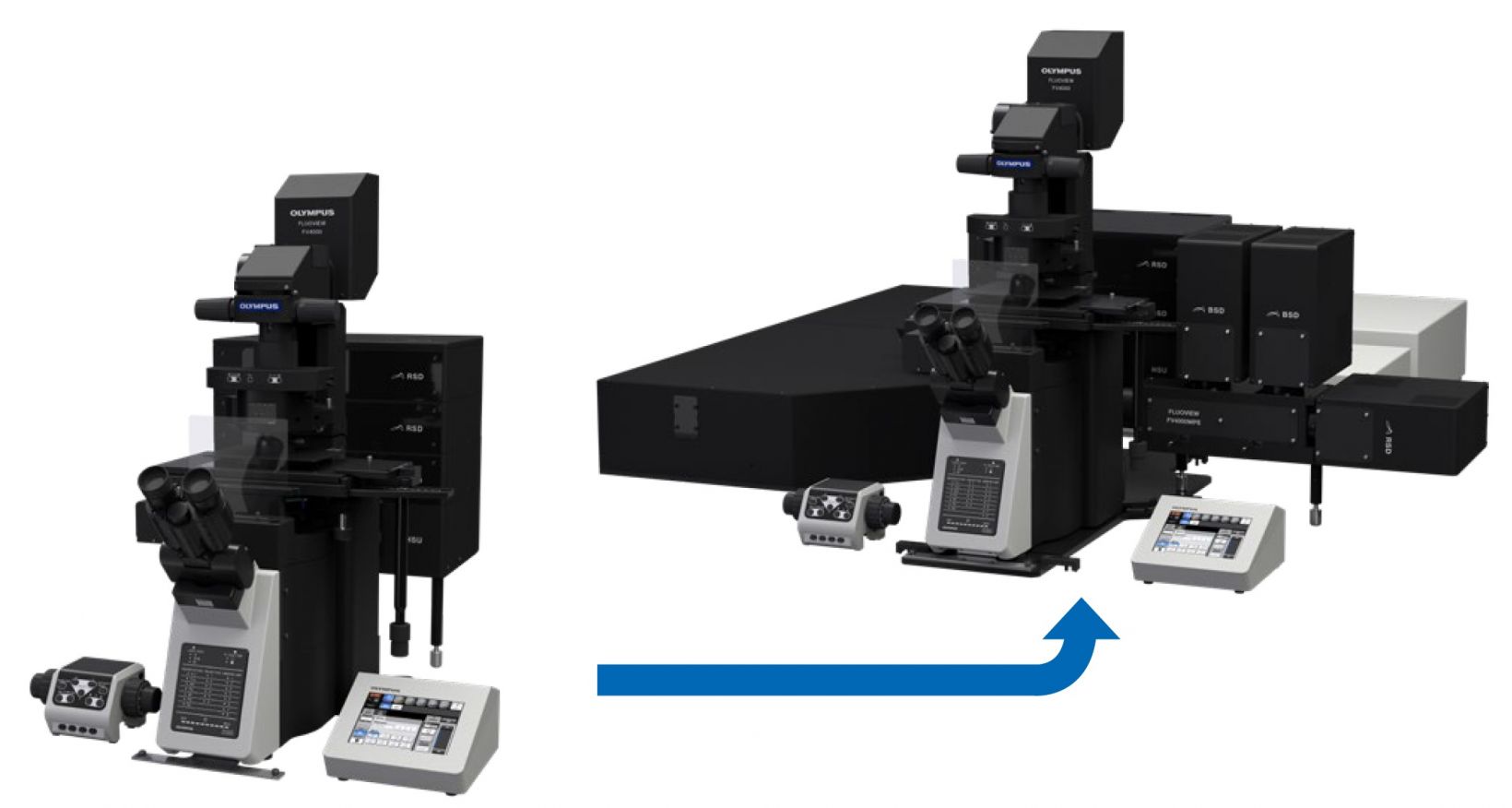
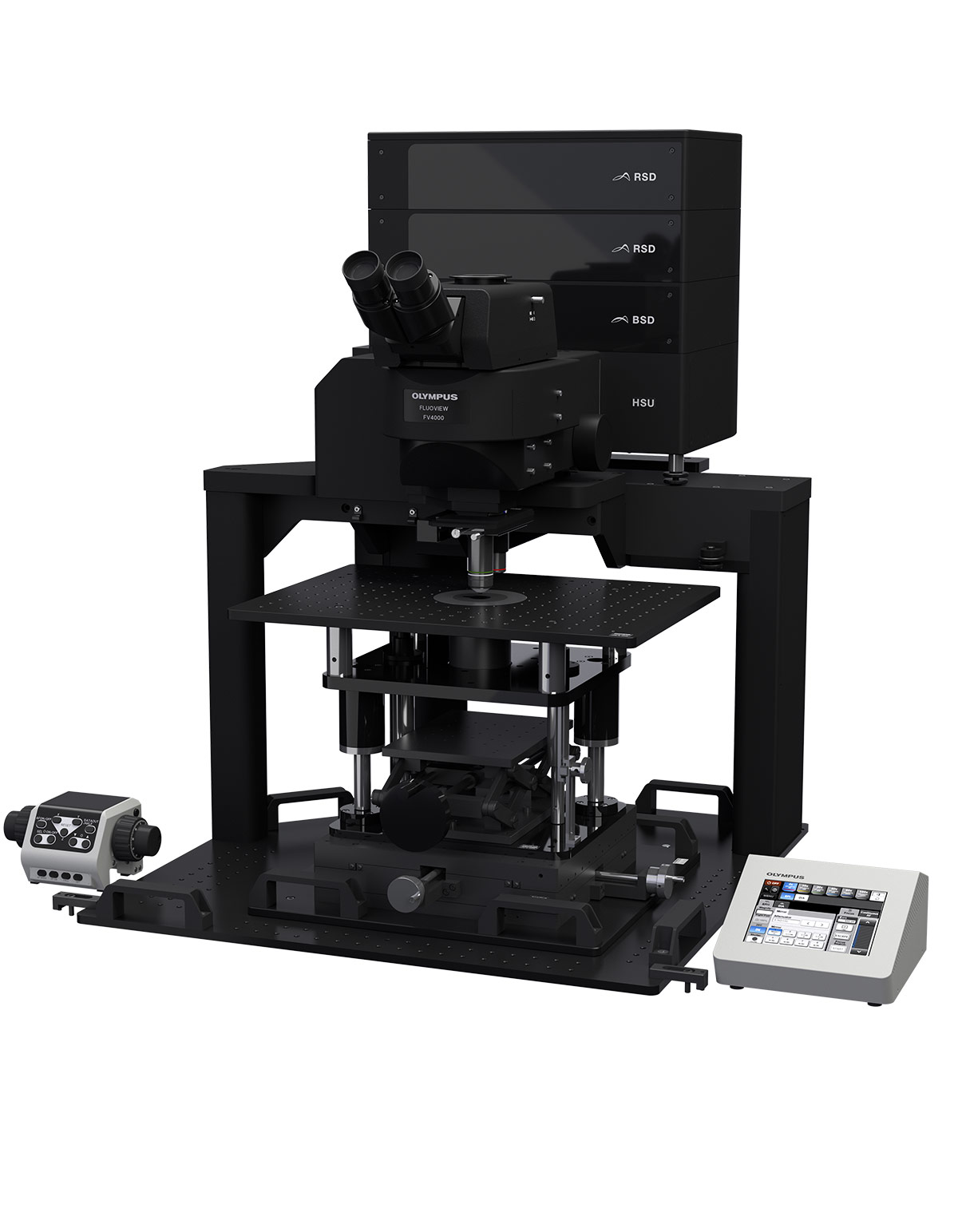
The FV4000 microscope is engineered to be modular, making it easy for you to configure the system based on your applications and budget. You can start with a standard FV4000 and easily upgrade to multiphoton imaging by adding the MPE module as your research changes.
| ||||||||
Need assistance? |
Specifications
| Scanner |
Galvanometer scanner
(normal imaging) | 64 × 64 to 4096 × 4096 pixels, 1 μs/pixel–1000 μs/pixel | |
|---|---|---|---|
|
Resonant scanner scanner
(high-speed imaging) | 512 × 512 pixels, 1024 × 1024 pixels | ||
| Field number (FN) | 20 | ||
| Spectral confocal detector | Detector | SilVIR detector (cooled SiPM, broadband type/red-shifted type) | |
| Maximum channels | Six channels | ||
| Spectral method | VPH, detectable wavelength range 400 nm–900 nm | ||
| Laser | VIS laser | 405 nm, 445 nm, 488 nm, 514 nm, 561 nm, 594 nm, 640 nm | |
| NIR laser | 685 nm, 730 nm, 785 nm | ||
| Laser power monitor | Built in | ||
| Image | High dynamic range photon counting (1G cps, 16-bit) | ||
