
Some of your favorite microscope images from October are covered in pollen. Who know something that makes us sneeze could be so pretty? Check out all the top images below featuring plants, diatoms, sea cucumbers, and more.
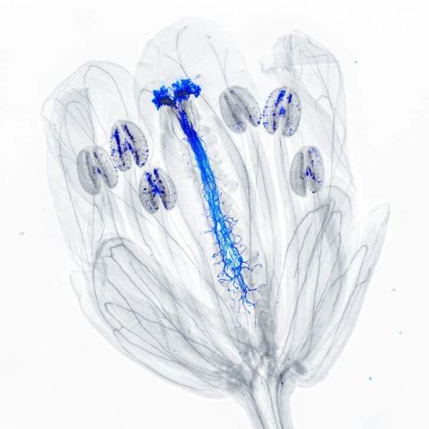
If you think this image looks familiar, you’re correct! What do you do when you have an already incredible image that has won an annual light microscopy imaging competition? You see how to make it even cooler looking! Jan Martinek’s image of an Arabidopsis thaliana flower won our Global Image of the Year competition in 2021, and this inversion has us in awe.
“In the image, the pollen tubes and pollen grains were stained with aniline blue, which binds to callose in their cell walls. In ultraviolet (UV) light, it has yellowish fluorescence glowing in the black background. Inverting the colors makes the tubes blue on a white background.”
Image and caption courtesy of Jan Martinek, global winner of the Evident Image of the Year 2021 competition. Captured on an Olympus AX70 microscope. To learn more about Jan’s prize-winning image, read our interview.
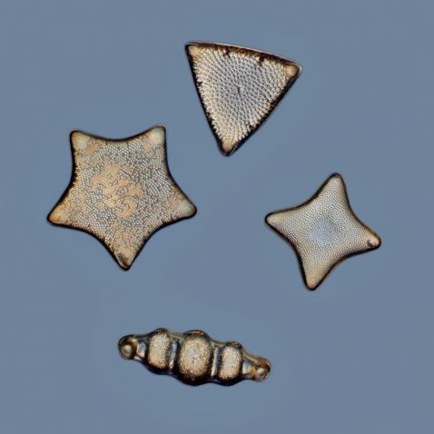
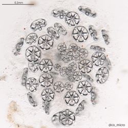
We love a good diatom arrangement, and so do you it seems! This arrangement has incredible detail in its small but mighty collection of a Biddulphia sp. on the bottom and three Triceratium sp.
Image and caption courtesy of @macro__cosmos. Captured on an Olympus IX73 inverted microscope.
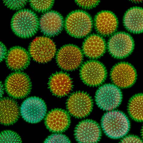
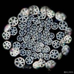
Achoo! We can’t help but sneeze looking at this stunning image of a pollen sample.
This image shows the “fluorescence microscopy of a Convolvulaceae sp. (likely Ipomoea) flower pollen. They are colloquially known as morning glory or the trumpet flower in China.”
Image and caption courtesy of @macro__cosmos. Captured on an Olympus IX73 inverted microscope.
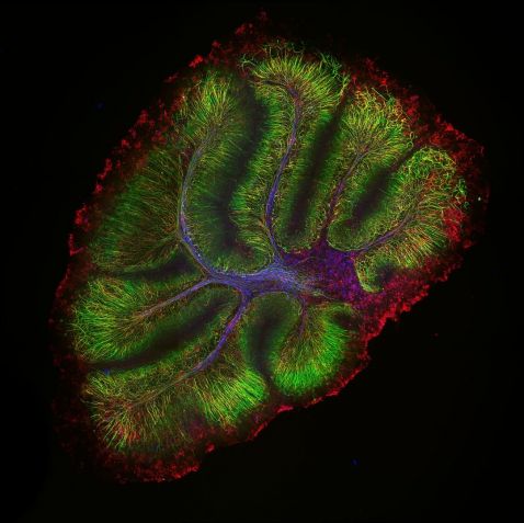
This month culminated in the launch of our FLUOVIEW™ FV4000 laser scanning confocal microscope with its SilVIR™ detector. This image of a mouse cerebellum demonstrates the imaging capabilities of this innovative system. Neurofilament-heavy chain (NFH) in green, myelin basic protein (MBP) in red, and glutathione S-transferase pi 1 (GSTpi) in blue.
Sample courtesy of Dr. Katherine Given, Principal Investigator, Neurobiology, University of Colorado Anschutz Medical Campus, Aurora, Colorado. Image captured on a FLUOVIEW FV4000 laser scanning confocal microscope using an UPLXAPO10X X Line™ objective.
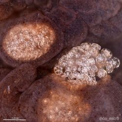
Polycheira rufescens, a genus of holothurians (sea cucumbers), is commonly found in the intertidal zone under rocks in Sagami Bay, Japan, where this microscopist often samples. They possess a unique internal skeleton, which is composed of thousands to millions of ossicles, or small calcareous elements beneath the skin.
Image courtesy of @co_micro. Captured on an Olympus BH2 microscope.
Bonus video! This is a bloody good one.
"The average human adult has more than five liters of blood in their body, which makes up 6% to 7% of the body weight! Blood delivers nutrients and oxygen to every organ, tissue, and cell of the living human body. Blood is composed of around 60% plasma, a fluid that mainly contains water, proteins, sugars, and fat. Red and white blood cells and platelets compose the 40% left.
The most common cells in the bloodstream are red blood cells, also called erythrocytes. The role of red blood cells is to transport oxygen from the lungs to the tissues with the help of a special protein called hemoglobin. Since red blood cells lack a nucleus, they have kind of a donut shape. This enables them to carry more hemoglobin and thus, more oxygen! Every second, around 2–4 million red blood cells are produced by the bone marrow and released into the circulatory system. There are around four to six million of these cells for each cubic millimeter of blood.
The white blood cells also have critical roles. They protect us from pathogens by engaging an immune response. The white blood cell from the video is most likely a macrophage, which crawl like an amoeba and eat harmful agents and damaged or old cells."
Video and caption courtesy of Chloé Savard. Captured on an Olympus BX53 microscope.
To see more images like these, be sure to follow us on Instagram at @evidentlifescience!
Want to share your own images? Visit our image submission site.
Related Content
Magnificent Microorganisms—Our Most Popular Microscope Images for September 2023
Post-Mortem Histology Artwork Brings Death to Life
Breadth of Beauty—Our Most Popular Microscope Images for August 2023
