What does death look like under the microscope? We spoke to Dr. Marianne Hamel, a forensic pathologist, to find out.
Marianne practices forensic pathology in Pennsylvania and New Jersey. With her creative partner Nikki Johnson, a forensic photographer in New York City, she is currently showing a collection of post-mortem histological images called Death Under Glass at the Stedman Gallery at Rutgers University in Camden, New Jersey.
These images offer a rare look into human anatomy after death—and are surprisingly beautiful. Each specimen is sliced extremely thin, then stained with an array of dyes highlighting particular elements of subcellular structure to illustrate the effects of natural disease, trauma, or drug abuse on the body. Some images in the collection are diagnostic and enable forensic pathologists to determine a cause of death; others are simply beautiful, illustrating the complex interplay of tissues in the human body. Beyond the gallery, Marianne also shares death investigation at high magnification on Instagram as @deathunderglass. We chatted with Marianne to learn more about her post-mortem histology artwork. The interview highlights her unique view of the world as a forensic pathologist and shares how microscopes bring these images about death to life. |  |
Q: Can you explain the role of a forensic pathologist and how it differs from other medical specialties?
Marianne: The most obvious difference between forensic pathologists and other medical specialties is that I don’t treat live patients. If I do, it's very rare.
As a forensic pathologist, my job is to determine the cause and manner of death for people who die violently, suspiciously, not under a doctor's care, or under any other circumstances that might warrant the attention of the local medical examiner. I do that by performing autopsies.
What I'm most interested in is figuring out the cause of death and the circumstances around it. It's like trying to figure out what happened in a play, but you only get to attend the last act. So, you have to be a good detective. It is a really good job for those who are a little bit nosey and who like to solve puzzles. You have to be able to work with your hands and be okay with having incomplete information.
The other good thing about the job is that if you hate being bored—and I’m very much that person—it's a great job. I get to do something different every day. Some days I do autopsies, review cold cases, look at histology, or do the occasional examination. I also review civil and criminal cases for attorneys. If you are a person who cannot do the same thing repeatedly, it's a great job. You also need a strong stomach, but that's a different issue.
Q: Who or what helped inspire you to become a forensic pathologist?
Marianne: When I was fourteen, I decided I wanted to be a pathologist. My sister decided she wanted to be a dentist when she was 12 and she became one, so we both picked a job and stuck with it.
And when I was 19, after my first year of college, I got an internship with the local medical examiner, Dr. Isidore Mihalakis, for part of Pennsylvania. He was great. He let me follow him around like a duckling all summer. We went to authorities, we went to court, we met with families, and we went to death scenes. I held test tubes, cleaned scalpel handles, and did whatever else he needed. It was a great experience.
Then I went to medical school. I walked in the first day, and I wanted to be a medical examiner. I walked out on the last day, and I still wanted to be a medical examiner. I never considered any other specialty. The end goal was always to become a board-certified forensic pathologist.
Q: How do you use microscopy in your everyday work?
Marianne: Most of the time, surgical pathologists, or people who read biopsies and tissue samples for a living, are largely looking to make a diagnosis. Most commonly, they are checking for cancer versus not cancer.
That's not commonly what I'm looking for. I’m looking for all sorts of things. Some diagnoses in forensic pathology can only be made by microscopy. For instance, myocarditis—or inflammation of the heart—can cause unexpected sudden death, leaving no other trace other than histologic evidence. Another example is antifreeze poisoning, which leaves fan-shaped crystals in the kidneys that you can see under polarized light. So, there are a number of diagnoses in forensic pathology that you must make through the microscope. I enjoy the microscope work quite a bit.
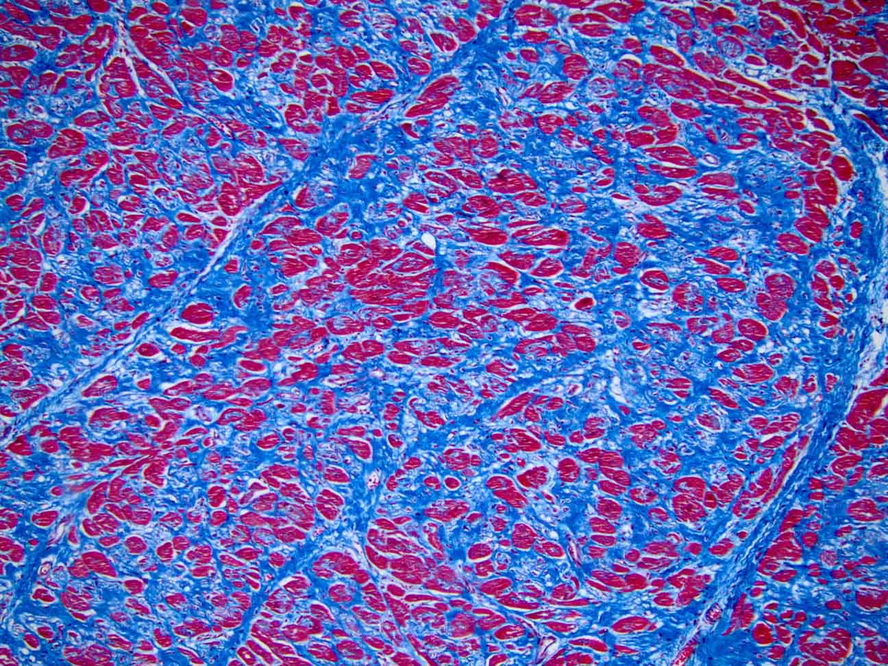
Section of a damaged human heart with a trichrome stain. Blue areas are scar tissue. Red areas are viable cells. Captured on an Olympus BX43 microscope with a DP26 camera. Image courtesy of Marianne Hamel.
Q: What types of microscopes do you commonly use in your lab?
Marianne: I read all my slides at home because my Olympus BX43 microscope is so good, and I use polarization. I rely on the histologist a lot to give me good special stains. Histology is a combination of art and science that does not get nearly enough recognition. If you have a good histologist, throw money and praise at them, and don’t let them go, because they're not making more histologists!
I use the Olympus cellSens™ software to process my images and save them. When we create the images for the show, we send them to a specialty printer in New York City and have them printed on aluminum.
Q: Can you describe a situation where a microscope was important to solving a case?
Marianne: I had a case not too long ago of a woman who had a drug problem. Her tests came back positive for methamphetamine. However, when I looked at a cross-section of one of her coronary arteries, I could see that it had dissected. There was a tear in the inner wall of her artery that had created a double channel of blood flow, and it was forcing shut the normal channel.
Her actual cause of death was not the methamphetamine toxicity. I would not have caught that if I hadn’t put that coronary artery through for microscopic examination.
Q: How has microscopy technology evolved over the course of your career, and how has it impacted your work?
Marianne: If you look at old microscope slides and in old textbooks, they're usually shot in black and white. They look like they're shot at a distance and through a fog bank. It’s not the fault of the microscopist. It’s just the limitations of the technology at the time.
Learning to read slides is like learning another language. The pathologist has an important role in that they interpret the tissue for other doctors so that they can make a diagnosis and offer appropriate treatment. To learn to do that, you need to read hundreds of thousands of slides with another pathologist who can help you learn that language.
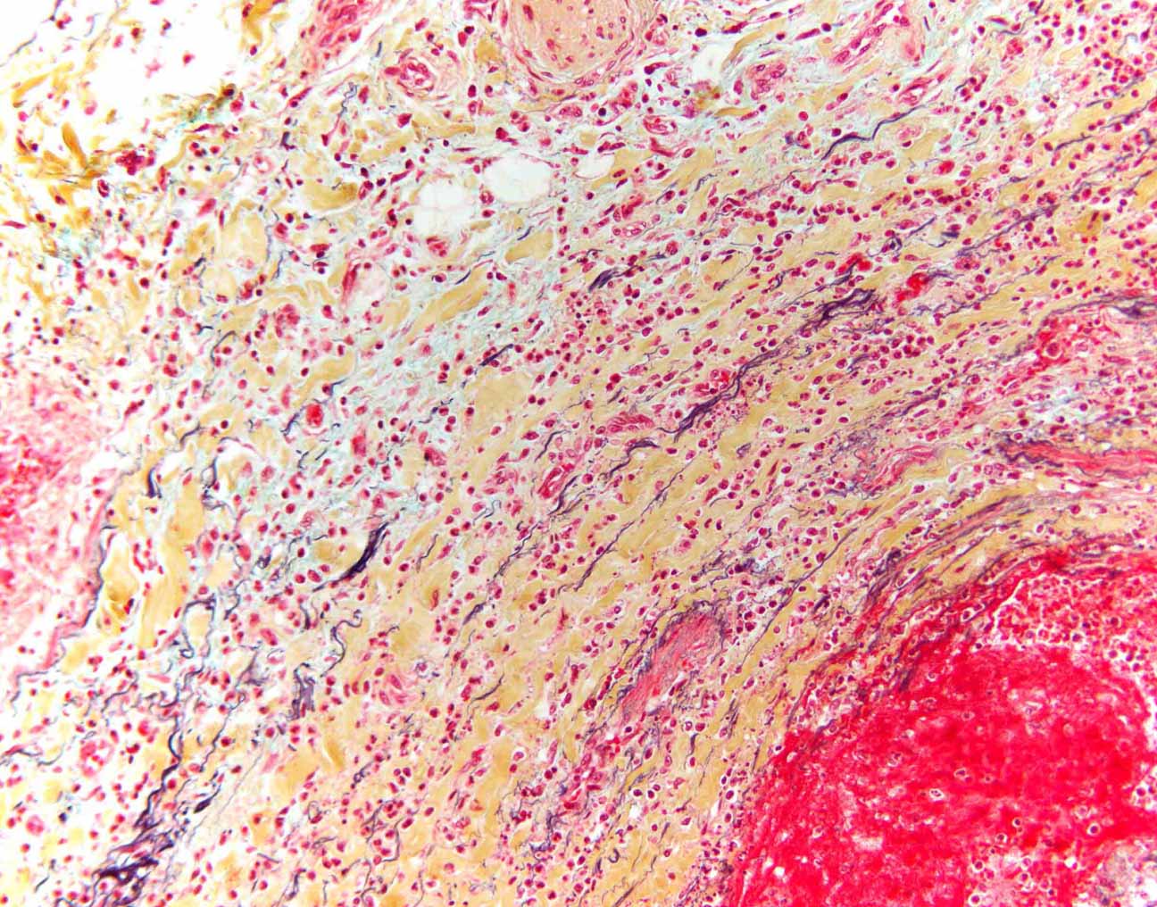
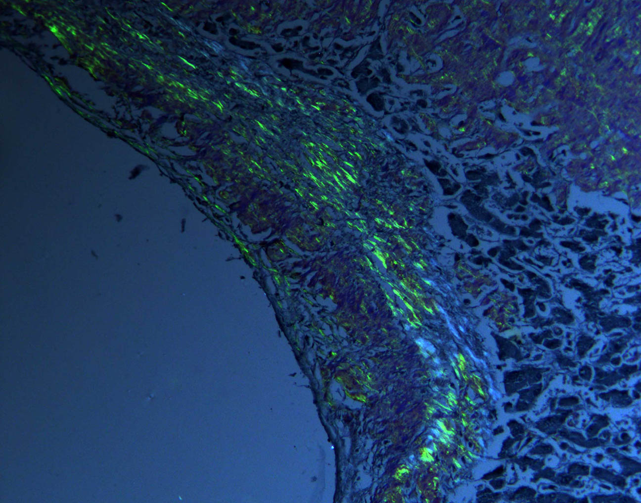
Section of normal human artery with Movat’s pentachrome stain, which uses five different dyes to highlight various aspects of subcellular anatomy. For instance, the elastic fibers in the artery wall, which give the blood vessel strength and resilience, appear black when treated with a pentachrome stain. Captured on an Olympus BX43 microscope with a DP26 camera. Image courtesy of Marianne Hamel.
Q: Do you collaborate with other professionals as part of your forensic pathology work?
Marianne: We run toxicology on almost every case. Particularly given the current fentanyl crisis, toxicology is incredibly important. Toxicologists have an extremely difficult job. They're basically building the airplane in the air as the fentanyl epidemic shifts and evolves into different things. By chance, my career started just as the opioid epidemic was taking off. I've been watching the epidemic change and evolve in real time for 12 years.
We sometimes call in an anthropologist. Every now and then we have a case that's either partially or totally skeletonized. I probably call in a bone specialist four or five times a year. They're incredibly important. As mentioned, I work with histologists frequently. Every case is a little bit different, and that's why I say I do something different every day. You have to modify your approach for every case.
Q: Have any of these collaborations extended into your art?
Marianne: To some extent, other pathologists have sent me slides and said, “This is really beautiful. You should look at this.” Sometimes a few of them have ended up in the show, and that's tremendous. Professional generosity on their part.
Q: When did you first realize that your work could also be considered art? How did you pursue that path?
Marianne: I was a trainee, and I showed some of the images through the microscope to Nikki Johnson, my creative collaborator. I said, “It would be so nice if you could show other people what this looked like.” She looked at them and she said, “If you can capture this, this is high resolution enough that we could blow it up and make actual art out of it.” And we did.
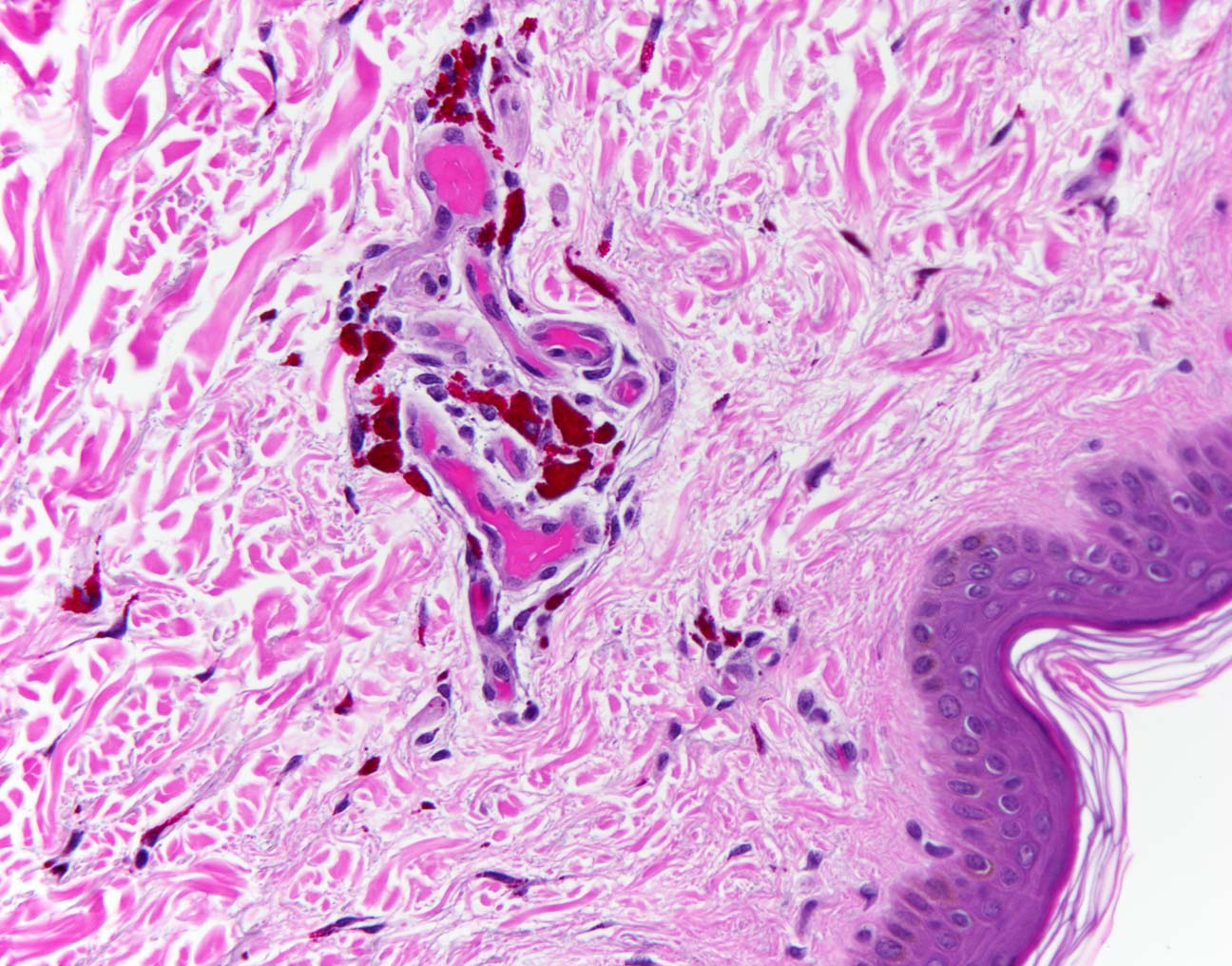
Section of human skin bearing a tattoo. Individual pigment granules are visible in the dermis, the deep layer of the skin, under high magnification. Other pigment granule colors that are identifiable under the microscope are black, blue, and gold. Captured on an Olympus BX43 microscope with a DP26 camera. Image courtesy of Marianne Hamel.
Q: What does it mean to you to have your work shown in a gallery?
Marianne: It's a little surreal to see your own work in a in a public place. I frequently forget that the gallery shows are public. People come up to me and say, “Hey, I saw your show.” I think to myself, “You did?” Anybody can walk in.
It is very gratifying to have an idea and see it come to fruition. Jake Foster, the manager at the Stedman Gallery, did a beautiful job of hanging the show. We hung the actual prints, and then high-resolution projections were placed on the back wall. The projections are of images we didn't print but still wanted to show. There are about 25 of those images, so it worked out really well.
Q: What messages do you hope to communicate to those who view your work?
Marianne: One thing that is exciting in the field is the idea of forensic genealogy, which has less to do with this project. Forensic genealogy is the practice of using genetic research to identify a suspect or victim in a critical case. For example, the perpetrator’s sample may not be in a database, but their relatives are. Genealogy can use family trees and family pedigrees to narrow down a suspect pool to a few people, or even one person. That was never possible before. If you weren't in the database, we were out of luck.
Forensic genealogy is incredibly powerful. It has broken a few cases lately that I believed to have been unbreakable. I'm very interested to see how that goes. I anticipate it knowing that I have some cold cases that were never solved. I fully expect to be called to testify in the next few years because they have been able to make an arrest.
Q: Has your art experience helped you communicate your microscopic findings in court or to non-scientific audiences?
Marianne: You get better at expressing things in nontechnical language. That's helpful. One thing I always try to explain to people is that histology is actually an artificial construct. Human tissue without stain is clear, so we actually created this image. It's not a natural image. Having to explain that to art audiences has given me better words to explain it to a jury so they understand what's going on.
When you testify, you're giving a tiny master class in this little corner of forensic science that we're talking about today. If your answer is more than three words long, you don't talk to the prosecutor or the defense attorney. You talk to the jury, and you can tell very quickly if you're getting through to them. If you say the carotid artery, the one you check your pulse for, and six of the jurors reach up and touch their neck, you know they're listening. Writing for a nontechnical art audience has improved my communication for the layperson who's on the jury.
Q: Do you have any advice for anyone who want to pursue a career in forensic pathology?
Marianne: Number one, if you want to be histologist, hurry up and go to school because we desperately need you. There are very few talented histologists available. With the demand for forensic services, we need them more and more for forensic pathologists. It’s a long road to get there, but I have found it very rewarding. You have to have a pretty thick skin. People are going to criticize your work. You're not going to get a thank you from your patients as some other specialists do. However, I find the work and intellectual discipline of it very rewarding.
Section of human tissue treated with a Congo Red special stain and viewed under polarized light, revealing the presence of amyloid. Amyloidosis, a disease in which proteins are inappropriately deposited in human tissue, glows apple-green under polarized light. Captured on an Olympus BX43 microscope with a DP26 camera. Image courtesy of Marianne Hamel.
Q: Any advice for forensic pathologists who might want to incorporate more art into their work?
Marianne: Send me your slides!
Q: Has sharing your work on Instagram driven more interest in forensic pathology?
Marianne: I've been managing the @deathunderglass account on Instagram for eight years now, and the amount of interest that people have in forensic science is incredibly deep. They are very loyal followers.
I'll give you an example. I made a reading list for my interns at the Montgomery County Office. A list of 15 forensic or forensic-related books that I thought were good quality. Not all of them were in popular culture. Some of them were textbooks—all kinds of things. I listed a couple of them on the account and mentioned that if you would like the entire list, please email me. I thought maybe 20 or 30 people would email me. I woke up the next morning to find 400 emails in my inbox, and I realized I had seriously underestimated the interest.
Q: Is there anything else that you would like to share about forensic science?
Marianne: There is a movement right now to sanitize our ideas about death. In some circles, we're supposed to never look at human anatomy and pretend that people don't die. I've spent my entire career predicated on the fact that we will all die. So far, I'm not wrong! The death rate is 100%.
It's important to have discussions about what we do in the morgue. Some older practitioners in the field believe that what we do in the morgue is supposed to be secret, and we're never supposed to talk about it, because it's dealing with dead bodies and dissection and things like that. I don't believe that at all.
I think that I provide a valuable public service. The people in my field work incredibly hard but get little recognition. I’m not just talking about forensic pathologists—but the autopsy technicians, the people who work in identification, and toxicologists.
They all work incredibly hard and deserve praise too. We can’t pretend that nobody has to stitch up the body after we're done, or nobody has to clean the morgue, like these are magical things that never happened. They work hard, they do their job really well, and I depend on them every day.
Related Content
Whole Slide Imaging and Artificial Intelligence (AI) Accelerates Lupus Nephritis Evaluation
Behind the Lens—Dr. Stephanie Shiers Creates Cover-Worthy Neuroscience Art